. 2014 May;14(5):679-86.
doi: 10.1002/mabi.201300486. Epub 2014 Jan 23.

Dual delivery of vascular endothelial growth factor and hepatocyte growth factor coacervate displays strong angiogenic effects

Affiliations

Dual delivery of vascular endothelial growth factor and hepatocyte growth factor coacervate displays strong angiogenic effects

Hassan K Awada et al. Macromol Biosci. 2014 May.

Abstract

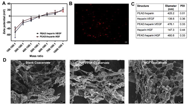
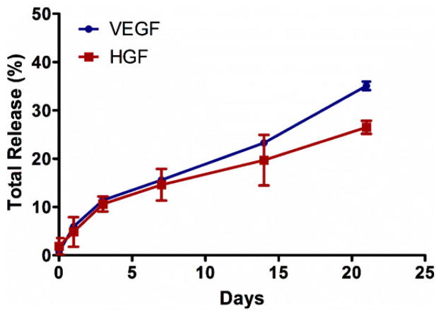
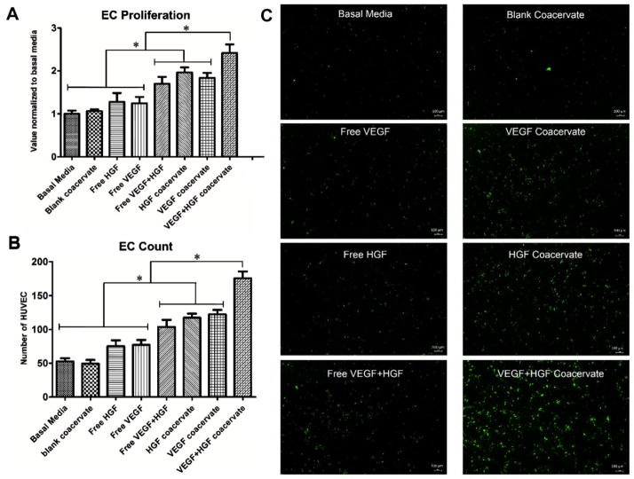
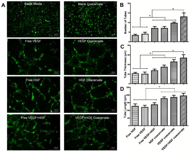
Controlled delivery of multiple growth factors (GFs) holds great potential for the clinical treatment of ischemic diseases and might be more therapeutically effective to reestablish vasculature than the provision of a single GF. Vascular endothelial growth factor (VEGF) and hepatocyte growth factor (HGF) are two potent angiogenic factors. However, due to rapid degradation and dilution in the body, their clinical potential will rely on an effective mode of delivery. A coacervate, composed of heparin and a biodegradable polycation, which protects GFs from proteolysis and potentiates their bioactivities, is developed. Here, the coacervate incorporates VEGF and HGF and sustains their release for at least three weeks. Their strong angiogenic effects on endothelial cell proliferation and tube formation in vitro are confirmed. Furthermore, it is demonstrated that coacervate-based delivery of these factors has stronger effects than free application of both factors and to coacervate delivery of each GF separately.

Keywords: angiogenesis; coacervate; drug delivery systems; growth factor; proteins.


Figures

Figure 1
Figure 1
A) Zeta potentials of VEGF and HGF coacervates were measured at different mass ratios of PEAD:heparin:GF by titrating heparin: GF solutions with PEAD. B) Spherical droplets of Rhodamine-labeled blank coacervates were imaged by fluorescence microscope. C) DLS measurements show the hydrodynamic diameters of heparin:GF, PEAD:heparin, and PEAD:heparin:GF particles. D) SEM images at low magnification (500×) show the ribbon-like structures and globular domains of blank coacervate, VEGF coacervate, and HGF coacervate.
Figure 2
Figure 2
Sustained in vitro release of VEGF and HGF from the coacervate for 3 weeks into DI water. VEGF and HGF were combined, then mixed with heparin followed by PEAD. After coacervate formed, tubes were centrifuged and supernatant was collected. GF amount was then quantified by sandwich ELISA at the specific time points. Bars indicate means ± SD.
Figure 3
Figure 3
Endothelial cell (EC) proliferation and live cell count assays. Treatment groups with 30 ng mL−1 concentration for each GF were applied to 3 wells per group with seeded HUVEC. A) One day after incubation, BrdU cell proliferation assay was performed and absorbance was recorded. Data is presented as a fold-change from the basal media. B) Live cell number was quantified after 3 days incubation over 0.67 mm2 fields in 3 wells per group. C) Microscope images of calcein-stained HUVEC in 4 mm2 fields. Bars indicate means ± SD. * p value <0.05.
Figure 4
Figure 4
Endothelial tube formation assay using 3D fibrin gels environment. HUVEC were seeded on bottom gel and specific treatment groups were added to top gel with 3 wells per group and incubated for 3 days. A) Microscope images of calcein-stained EC tubes formed in different groups. B) Number of EC tubes, C) tube thickness, and D) tube length were quantified by microscope imaging analysis software in 0.67 mm2 fields. Bars indicate means ± SD. * p value <0.05.

References

    1. Tayalia P, Mooney DJ. Adv Mater. 2009;21:3269. - PubMed
    1. Li WW, Talcott KE, Zhai AW, Kruger EA, Li VW. Adv Skin Wound Care. 2005;18:491. - PubMed
    1. Chu H, Wang Y. Ther Delivery. 2012;3:693. - PMC - PubMed
    1. Chen FM, Zhang M, Wu ZF. Biomaterials. 2010;31:6279. - PubMed
    1. Risau W. Nature. 1997;386:671. - PubMed

Publication types

MeSH terms

Substances