Review
. 2014 Jan;55(1):5-10; quiz 11.
doi: 10.11622/smedj.2014003.

Lipoma arborescens

Affiliations
Review

Lipoma arborescens

Sarat Kumar Sanamandra et al. Singapore Med J. 2014 Jan.

Abstract

Lipoma arborescens is a chronic, slowly progressive intra-articular lesion characterised by villous lipomatous proliferation of the synovium, usually involving the suprapatellar pouch of the knee joint. It is an uncommon cause of intra-articular masses that presents as slowly progressive painless swelling of the joint, which persists for many years and is accompanied by intermittent effusions. We highlight this condition to raise awareness of its clinical spectrum and imaging features, so that early diagnosis and appropriate treatment can be given, and misinterpretation of this condition as other more complex intra-articular masses is avoided. This pictorial essay aims to provide a brief yet comprehensive review of the clinical features, distribution, morphological types and imaging characteristics of lipoma arborescens, including its common differential diagnoses and management.


Figures

Fig. 1
Fig. 1
Photomicrographs of extra-articular lipoma arborescens show several papillaroid structures (arrows) with synovial lining cells (arrowheads), which contain a stroma that exhibits increased mature adipose tissue (stars) [Haematoxylin & eosin (a) × 2; (b) × 4].
Fig. 2
Fig. 2
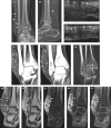
A 43-year-old woman with slowly progressive painless swelling along the lateral aspect of the right ankle joint. (a) Frontal and (b) lateral view radiographs of the ankle typically show lobulated soft tissue opacity on the lateral aspect that contains a few internal faint lucent foci (arrows), compatible with underlying prominent fatty projections. US images of the lesion (c) without and (d) with probe compression demonstrate soft consistency of the lesion (as opposed to the firm and noncompressible nature of pigmented villonodular synovitis [PVNS]), containing heterogeneous hyperechoic nodular areas (arrows) that are similar in echotexture with adjacent subcutaneous fat (star). Coronal reformatted CT images in (e & f) soft tissue and (g) bone windows reveal a lobulated mass at the lateral aspect of the ankle, containing several fat density nodular foci (arrows) interspersed in fluid or soft tissue density component (arrowheads). Note that there is an absence of bony or articular surface erosions in Fig. 2g, an important clue in the differential diagnosis for PVNS or gouty arthropathy. Coronal (h) T1, (i) T2 and (j) T2 fat-saturated MR images of the ankle display several nodular and villous fatty masses (arrows) within the lesion that are surrounded by a trace of effusion and hypertrophied synovium (arrowheads). (k) Coronal gradient image shows an absence of susceptibility artifact, which would indicate the lack of haemorrhagic products in the lesion. (l) Post-contrast T1 fat-saturated MR image of the lesion shows several non-enhancing fatty masses (arrows) with enhancing surrounding soft tissue/thickened synovium (arrowheads).
Fig. 3
Fig. 3
A 65-year-old woman with longstanding intermittent bilateral knee joint swelling and discomfort, which is more severe on the right side. (a) Frontal and (b) lateral view radiographs of the right knee joint show nonspecific suprapatellar fullness and a soft tissue, mass-like opacity (arrows) associated with marked background degenerative changes (arrowheads). Coronal (c) T1 and (d) T2 fat-saturated MR images of the right knee joint show the typical features of lipoma arborescens (arrows) with background degenerative changes in the form of marginal osteophytes (arrowheads), compatible with the secondary form of lipoma arborescens. (e) Coronal T1 MR image of the left knee joint also shows several intra-articular fatty villous projections, although to a lesser extent, in the suprapatellar bursa (arrows) associated with background degenerative changes (arrowhead), which is an example of multifocal secondary lipoma arborescens.
Fig. 4
Fig. 4
A 13-year-old boy with insidious onset of painless progressive left knee swelling that persisted for three years. (a) Sagittal T1 and (b & c) axial T2 fat-saturated MR images of the left knee show diffuse villous and frond-like intra-articular fatty masses (arrows) in the suprapatellar bursa with associated marked joint effusion (star). (d & e) Coronal post-contrast T1 fat-saturated images show non-enhancing fatty masses (arrow) and an enhancing overlying thickened synovium (arrowheads). The younger age of onset, absence of background degenerative change or meniscal/ligament tears in this patient are indicative of a primary form of lipoma arborescens. (f & g) Arthroscopic images of the affected knee joint demonstrate several villous and frond-like fatty projections (arrows) arising from the synovial lining.
Fig. 5
Fig. 5
A 63-year-old woman with a longstanding history of intermittent swelling and pain in the right knee. Coronal and axial (a & c) T1 and (b & d) T2 fat-saturated MR images of the right knee show several tiny intra-articular fatty villous projections (arrows) scattered throughout the suprapatellar bursa, compatible with diffuse lipoma arborescens associated with background degenerative changes and large effusion (star).
Fig. 6
Fig. 6
A 61-year-old man with chronic left knee pain and intermittent swelling. (a & b) Coronal and axial T1 and (c) axial T2 fat-saturated MR images of the left knee show a nodular frond-like intra-articular fatty mass (arrow) at the lateral aspect of the suprapatellar bursa with surrounding effusion (star), consistent with nodular lipoma arborescens. (d) Axial post-contrast T1 fat-saturated MR image also shows a typical non-enhancing fatty mass (arrow) and an enhancing overlying thickened synovium (arrowhead), which were confirmed on subsequent arthroscopy (e).
Fig. 7
Fig. 7
A 59-year-old woman with longstanding intermittent joint swelling and low-grade pain in the left knee. (a, b & d) Sagittal, axial and coronal T1; and (c) axial T2 fat-saturated MR images of the left knee show an intra-articular frond-like fatty mass (arrow) in addition to several scattered fatty villous projections (arrowheads) in the suprapatellar bursa, consistent with a mixed morphologic pattern of lipoma arborescens. Arthroscopy of the concerned knee joint correspondingly demonstrates (e) a nodular fatty mass, and (f) a few scattered villous projections in the suprapatellar fossa.
Fig. 8
Fig. 8
A 45-year-old woman with longstanding right shoulder joint discomfort. (a) Axial T1 and (b) coronal T2 fat-saturated MR images of the shoulder joint show a lobulated fatty mass in the subacromial-subdeltoid bursa (arrows) associated with a small effusion (star), consistent with extra-articular lipoma arborescens.

References

    1. Hallel T, Lew S, Bansal M. Villous lipomatous proliferation of the synovial membrane (lipoma arborescens) J Bone Joint Surg Am. 1988;70:264–70. - PubMed
    1. Yan CH, Wong JWK, Yip DK. Bilateral knee lipoma arborescens: a case report. J Orthop Surg (Hong Kong) 2008;16:107–10. - PubMed
    1. Ryu KN, Jaovishida S, Schweitzer M, Motta AO, Resnick D. MR imaging of lipoma arborescens of the knee joint. Am J Roentgenol. 1996;167:1229–32. - PubMed
    1. Vilanova JC, Barceló J, Villalón M, et al. MR Imaging of Lipoma arborescens and the associated lesions. Skeletal Radiol. 2003;32:504–9. - PubMed
    1. Sheldon PJ, Forrester DM, Learch TJ. Imaging of intraarticular masses. Radiographics. 2005;25:105–19. - PubMed

LinkOut - more resources