. 2014 Mar 1;74(5):1576-87.
doi: 10.1158/0008-5472.CAN-13-1656. Epub 2014 Jan 22.

Mesenchymal stem cells use IDO to regulate immunity in tumor microenvironment

Affiliations

Mesenchymal stem cells use IDO to regulate immunity in tumor microenvironment

Weifang Ling et al. Cancer Res. .

Abstract

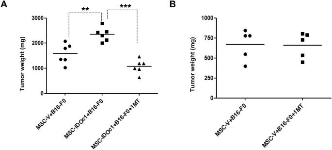
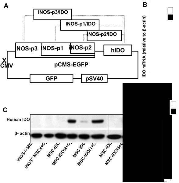
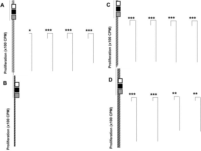
Mesenchymal stem cells (MSC) are present in most, if not all, tissues and are believed to contribute to tissue regeneration and the tissue immune microenvironment. Murine MSCs exert immunosuppressive effects through production of inducible nitric oxide synthase (iNOS), whereas human MSCs use indoleamine 2,3-dioxygenase (IDO). Thus, studies of MSC-mediated immunomodulation in mice may not be informative in the setting of human disease, although this critical difference has been mainly ignored. To address this issue, we established a novel humanized system to model human MSCs, using murine iNOS(-/-) MSCs that constitutively or inducibly express an ectopic human IDO gene. In this system, inducible IDO expression is driven by a mouse iNOS promoter that can be activated by inflammatory cytokine stimulation in a similar fashion as the human IDO promoter. These IDO-expressing humanized MSCs (MSC-IDO) were capable of suppressing T-lymphocyte proliferation in vitro. In melanoma and lymphoma tumor models, MSC-IDO promoted tumor growth in vivo, an effect that was reversed by the IDO inhibitor 1-methyl-tryptophan. We found that MSC-IDO dramatically reduced both tumor-infiltrating CD8(+) T cells and B cells. Our findings offer an important new line of evidence that interventional targeting of IDO activity could be used to restore tumor immunity in humans, by relieving IDO-mediated immune suppression of MSCs in the tumor microenvironment as well as in tumor cells themselves.


Figures

Figure 1
Figure 1. Constitutive expression of human IDO in mouse MSCs deficient in iNOS
(A) Design of the human IDO constitutive expression construct. Human IDO (hIDO) cDNA fragment was inserted into the pcDNA3.0 plasmid under control of the CMV promoter. Successful transfectants were selected using Neomycin. (B) Human IDO expression by MSCs transfected with IDO constitutive expression vector. Vectors with or without the human IDO gene construct were transfected into mouse iNOS−/− MSCs. Cells were harvested after two days in culture, total mRNA was extracted, and IDO message was assayed by real-time PCR, normalized to β-actin mRNA (defined as 1000 arbitrary units). MSC-IDOc1 and MSC-IDOc2: two representative human IDO constitutive expression clones. MSC-V: MSCs transfected with pcDNA3.0 vector only (negative control). (C) Human IDO protein was determined by western blotting analysis. MSCs were cultured for two days, harvested, and total cell lysates assayed for human IDO. MSC-V served as a negative control; human MSCs stimulated with inflammatory cytokines (TNFα, IL-1β, IFNγ; 20 ng/ml each) served as positive control. Results are representative of three independent experiments.
Figure 2
Figure 2. MSCs constitutively expressing human IDO inhibit the proliferation of both human and mouse lymphocytes
(A-B) Effect of MSC-IDOc on lymphocyte proliferation. MSC-IDOc were co-cultured with human PBMCs stimulated with OKT3 antibody (A) or mouse splenocytes stimulated with anti-CD3 and anti-CD28 (B) at a MSC-to-lymphocyte ratio of 1:10. Cell proliferation was assessed by H-thymidine incorporation after 48 hr. (C-D) 1-MT reversed the immunosuppressive effect mediated by MSC-IDOc. MSCs were co-cultured with mouse splenocytes at graded ratios of MSCs-to -splenocytes with or without 1-MT (0.5 mM), and cell proliferation was assessed by H-thymidine incorporation after 48 hr (C). MSC transfected with pcDNA3.0 empty vector (MSC-V) were not immunosuppressive (D). Values represent means ± SD from a representative of three experiments; *p < 0.05, **p < 0.01, ***p <0.001.
Figure 3
Figure 3. Human IDO constitutively expressed by MSCs Promotes Tumor Growth
(A-B) Wild type C57BL/6 mice were injected i.m. in the thigh with B16-F0 melanoma cells (0.5 × 10), and then administered intraperitoneally with MSC-IDOc or vector-transfectants (1 × 10 per mouse) every 2 days, with or without 1-MT (2 mg/ml) supplementation in drinking water. After 14 days, mice were sacrificed and resultant tumors were excised and weighed (A). 1-MT had no effect on mice treated with vector-transfectants (MSC-V) (B)
Figure 4
Figure 4. Inducible expression of human IDO in mouse MSCs deficient in iNOS
(A) Design of the human IDO inducible expression constructs. The original CMV promoter of the pCMS-EGFP vector was removed. Mouse iNOS promoter and human IDO cDNA fragments were then inserted into the vector. Three different versions of mouse iNOS promoter were incorporated: mouse iNOS promoter p2 (1750 base pairs upstream from ATG) was the essential part of the entire promoter; mouse iNOS promoter p1 was extended from p2 (1750+1650 base pairs); mouse iNOS promoter p3 was further extended from p1 (1750+1650+1650 base pairs). GFP was included as a reporter. (B) Each of the three inducible IDO expression constructs (iNOS-p1/IDO, iNOS-p2/IDO, iNOS-p3/IDO) were transfected into mouse iNOS−/− MSCs to generate three different MSC transfectants with inducible human IDO (hIDO) expression (MSC-IDOi1, MSC-IDOi2 and MSC-IDOi3, respectively). These transfectants were cultured with or without inflammatory cytokines (TNFα, IL-1β, IFNγ; 20 ng/ml each) for 24 hr, cells were harvested, and total mRNA was extracted. human IDO message was assayed by real-time PCR, normalized to β-actin mRNA (defined as 1000 arbitrary units). Non-transfected iNOS−/− MSCs served as negative control. (C) The same inducible hIDO-expressing MSC transfectants were assayed for hIDO expression at the protein level by western blotting analysis after culture, as in (B). Results are representative of three experiments. (D) To determine IDO enzyme activity, MSCs were cultured with or without inflammatory cytokine (TNFα, IL-1β, IFNγ; 20 ng/ml each) stimulation for 24 hr. IDO activity was assayed by spectrophotometric detection of the tryptophan metabolite, kynurenine, a product of IDO catabolism.
Figure 5
Figure 5. MSCs with inducible human IDO expression inhibit proliferation of both mouse and human lymphocytes
(A-D) Effects of MSC-IDOi on lymphocyte proliferation. MSCs were co-cultured with freshly-isolated mouse splenocytes at graded ratios of MSCs-to-splenocytes in the presence of soluble anti-CD3 and anti-CD28, and IFNγ, TNFα, and IL-1β (10 ng/ml each), with or without 1-MT (0.5 mM). After 48 hr, proliferation was assessed by H thymidine incorporation (A). Negative control mouse iNOS−/− MSCs were not immunosuppressive under identical conditions (B).The effects of wild type MSCs were similarly tested (C).The effects of MSC-IDOi on human lymphocyte proliferation was tested using human T cell blasts (D).
Figure 6
Figure 6. MSCs with inducible expression of human IDO promote tumor growth through modulation of immune cells
(A-C) Effects of MSCs with inducible human IDO (hIDO) expression on tumor growth. Wild type C57BL/6 mice or iNOS−/− mice were injected i.m. in the thigh with B16-F0 melanoma cells (0.5 × 10) (A, B) or EL4 lymphoma cells (0.5 × 10) (C), and then administered i.p. with MSCs having inducible hIDO-expression (MSC-IDOi1), or constitutive hIDO-expression (MSC-IDOc1; positive control), wild type (wt) MSCs, or negative control MSCs (MSC-control) (1 × 10 cells) every 2 days, with or without 1-MT (2 mg/ml) supplementation in drinking water. After 14 days, mice were sacrificed and resultant tumors were excised and weighed. (D) Distribution of immune cell subpopulations within tumor tissue. Cells were harvested from the tumors in (A), stained for the indicated markers, and analyzed by flow cytometry. The number of CD3+ T cells, CD4+ T cells, CD8+ T cells, CD4+ Foxp3+ T cells (regulatory T cells, Tregs), NKG2D+ cells (NK cells) and CD19+ cells (B cells) as a percentage of total cells within the tumor are shown. *p <0.05, **p < 0.01, ***p <0.001.
Figure 7
Figure 7. Tumor promotion by human IDO-transfected MSCs is abolished in immunodeficient mice
Rag1−/−, β2M−/− or CIITA−/− mice were treated as described in (Fig. 6A) and tumor growth measured. MSCs expressing either constitutive or inducible human IDO did not promote tumor growth in these mice.

References

    1. Aggarwal S, Pittenger MF. Human mesenchymal stem cells modulate allogeneic immune cell responses. Blood. 2005;105(4):1815–22. - PubMed
    1. Gebler A, Zabel O, Seliger B. The immunomodulatory capacity of mesenchymal stem cells. Trends Mol Med. 2011 - PubMed
    1. Zappia E, Casazza S, Pedemonte E, Benvenuto F, Bonanni I, Gerdoni E, et al. Mesenchymal stem cells ameliorate experimental autoimmune encephalomyelitis inducing T-cell anergy. Blood. 2005;106(5):1755–61. - PubMed
    1. Nauta AJ, Fibbe WE. Immunomodulatory properties of mesenchymal stromal cells. Blood. 2007;110(10):3499–506. - PubMed
    1. Ren G, Zhang L, Zhao X, Xu G, Zhang Y, Roberts AI, et al. Mesenchymal stem cell-mediated immunosuppression occurs via concerted action of chemokines and nitric oxide. Cell Stem Cell. 2008;2(2):141–50. - PubMed

Publication types

MeSH terms

Substances