

Progression of brain atrophy in the early stages of Parkinson's disease: a longitudinal tensor-based morphometry study in de novo patients without cognitive impairment
- PMID: 24453162
- PMCID: PMC6868950
- DOI: 10.1002/hbm.22449
Progression of brain atrophy in the early stages of Parkinson's disease: a longitudinal tensor-based morphometry study in de novo patients without cognitive impairment
Abstract
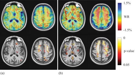
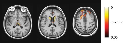
The presence of brain atrophy and its progression in early Parkinson's disease (PD) are still a matter of debate, particularly in patients without cognitive impairment. The aim of this longitudinal study was to assess whether PD patients who remain cognitively intact develop progressive atrophic changes in the early stages of the disease. For this purpose, we employed high-resolution T1-weighted MR imaging to compare 22 drug-naïve de novo PD patients without cognitive impairment to 17 age-matched control subjects, both at baseline and at three-year follow-up. We used tensor-based morphometry to explore the presence of atrophic changes at baseline and to compute yearly atrophy rates, after which we performed voxel-wise group comparisons using threshold-free cluster enhancement. At baseline, we did not observe significant differences in regional atrophy in PD patients with respect to control subjects. In contrast, PD patients showed significantly higher yearly atrophy rates in the prefrontal cortex, anterior cingulum, caudate nucleus, and thalamus when compared to control subjects. Our results indicate that even cognitively preserved PD patients show progressive cortical and subcortical atrophic changes in regions related to cognitive functions and that these changes are already detectable in the early stages of the disease.
Keywords: TBM; brain atrophy; cognitive status; de novo PD; longitudinal study.
Copyright © 2014 Wiley Periodicals, Inc.
Figures
Similar articles
-
Longitudinal brain atrophy distribution in advanced Parkinson's disease: What makes the difference in "cognitive status" converters?Hum Brain Mapp. 2020 Apr 15;41(6):1416-1434. doi: 10.1002/hbm.24884. Epub 2019 Dec 2. Hum Brain Mapp. 2020. PMID: 31789477 Free PMC article.
-
Progression of subcortical atrophy in mild Parkinson's disease and its impact on cognition.Eur J Neurol. 2017 Feb;24(2):341-348. doi: 10.1111/ene.13205. Epub 2016 Dec 10. Eur J Neurol. 2017. PMID: 27943468
-
Changes of brain structure in Parkinson's disease patients with mild cognitive impairment analyzed via VBM technology.Neurosci Lett. 2017 Sep 29;658:121-132. doi: 10.1016/j.neulet.2017.08.028. Epub 2017 Aug 18. Neurosci Lett. 2017. PMID: 28823894
-
Progression of grey and white matter brain damage in Parkinson's disease: a critical review of structural MRI literature.J Neurol. 2021 Sep;268(9):3144-3179. doi: 10.1007/s00415-020-09863-8. Epub 2020 May 6. J Neurol. 2021. PMID: 32378035 Review.
-
MRI and cognitive impairment in Parkinson's disease.Mov Disord. 2009;24 Suppl 2:S748-53. doi: 10.1002/mds.22670. Mov Disord. 2009. PMID: 19877242 Review.
Cited by
-
Shared and distinct cortical morphometric alterations in five neuropsychiatric symptoms of Parkinson's disease.Transl Psychiatry. 2024 Aug 30;14(1):347. doi: 10.1038/s41398-024-03070-z. Transl Psychiatry. 2024. PMID: 39214962 Free PMC article.
-
Subthalamic Beta Activity in Parkinson's Disease May Be Linked to Dorsal Striatum Gray Matter Volume and Prefrontal Cortical Thickness: A Pilot Study.Front Neurol. 2022 Mar 23;13:799696. doi: 10.3389/fneur.2022.799696. eCollection 2022. Front Neurol. 2022. PMID: 35401426 Free PMC article.
-
How should we be using biomarkers in trials of disease modification in Parkinson's disease?Brain. 2023 Dec 1;146(12):4845-4869. doi: 10.1093/brain/awad265. Brain. 2023. PMID: 37536279 Free PMC article.
-
Progression of brain atrophy in spinocerebellar ataxia type 2: a longitudinal tensor-based morphometry study.PLoS One. 2014 Feb 25;9(2):e89410. doi: 10.1371/journal.pone.0089410. eCollection 2014. PLoS One. 2014. PMID: 24586758 Free PMC article.
-
Dopamine selectively remediates 'model-based' reward learning: a computational approach.Brain. 2016 Feb;139(Pt 2):355-64. doi: 10.1093/brain/awv347. Epub 2015 Dec 17. Brain. 2016. PMID: 26685155 Free PMC article.
References
-
- Aarsland D, Bronnick K, Williams‐Gray C, Weintraub D, Marder K, Kulisevsky J, Burn D, Barone P, Pagonabarraga J, Allcock L, Santangelo G, Foltynie T, Janvin C, Larsen JP, Barker RA, Emre M (2010): Mild cognitive impairment in Parkinson's disease: A multicenter pooled analysis. Neurology 75:1062–1069. - PMC - PubMed
-
- Albin RL, Young AB, Penney JB (1989): The functional anatomy of basal ganglia disorders. Trends Neurosci 12:366–375. - PubMed
-
- Allen JS, Bruss J, Brown CK, Damasio H (2005): Normal neuroanatomical variation due to age: The major lobes and a parcellation of the temporal region. Neurobiol Aging 26:1245–1260. - PubMed
-
- American Psychiatric Association . 2000. Diagnostic and Statistical Manual of Mental Disorders, fourth edition, Text Revision. Washington, DC: American Psychiatric Publication, 943 p.
-
- Antonini A, De Notaris R, Benti R, De Gaspari D, Pezzoli G (2001): Perfusion ECD/SPECT in the characterization of cognitive deficits in Parkinson's disease. Neurol Sci 22:45–46. - PubMed
MeSH terms
LinkOut - more resources
Full Text Sources
Other Literature Sources
Medical
