. 2014 Mar;164(3):1175-90.
doi: 10.1104/pp.113.233866. Epub 2014 Jan 22.

Plastidial NAD-dependent malate dehydrogenase is critical for embryo development and heterotrophic metabolism in Arabidopsis

Affiliations

Plastidial NAD-dependent malate dehydrogenase is critical for embryo development and heterotrophic metabolism in Arabidopsis

Seraina Beeler et al. Plant Physiol. 2014 Mar.

Abstract

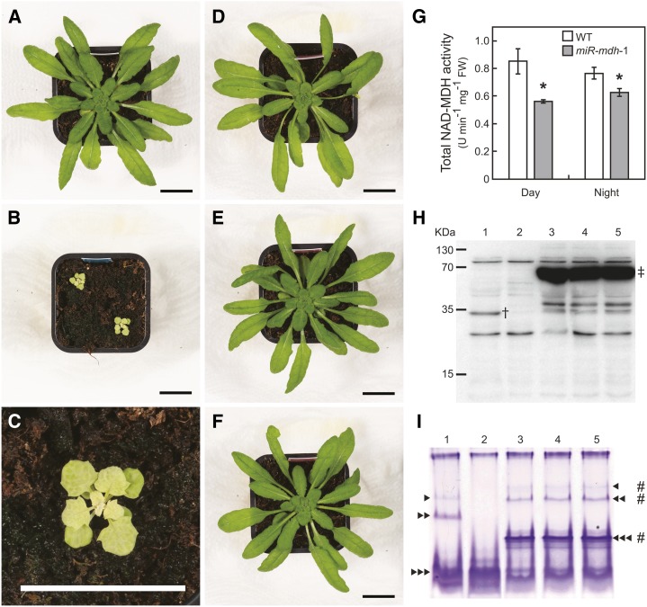
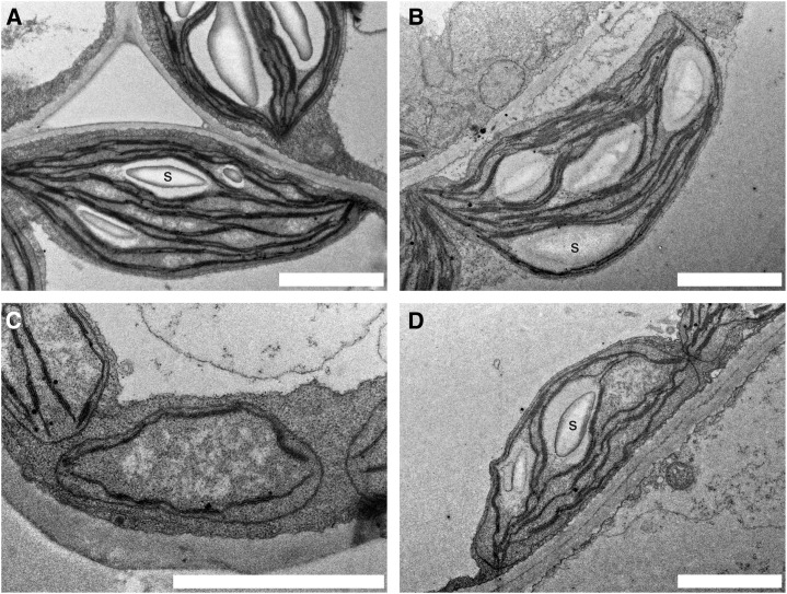
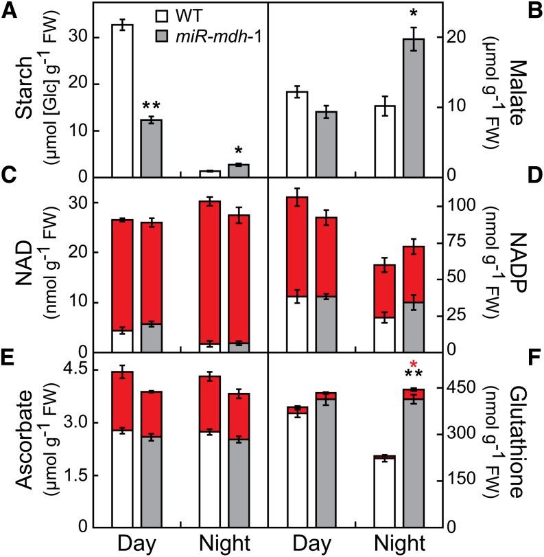
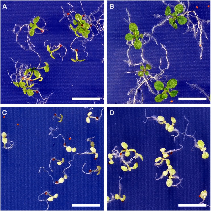
In illuminated chloroplasts, one mechanism involved in reduction-oxidation (redox) homeostasis is the malate-oxaloacetate (OAA) shuttle. Excess electrons from photosynthetic electron transport in the form of nicotinamide adenine dinucleotide phosphate, reduced are used by NADP-dependent malate dehydrogenase (MDH) to reduce OAA to malate, thus regenerating the electron acceptor NADP. NADP-MDH is a strictly redox-regulated, light-activated enzyme that is inactive in the dark. In the dark or in nonphotosynthetic tissues, the malate-OAA shuttle was proposed to be mediated by the constitutively active plastidial NAD-specific MDH isoform (pdNAD-MDH), but evidence is scarce. Here, we reveal the critical role of pdNAD-MDH in Arabidopsis (Arabidopsis thaliana) plants. A pdnad-mdh null mutation is embryo lethal. Plants with reduced pdNAD-MDH levels by means of artificial microRNA (miR-mdh-1) are viable, but dark metabolism is altered as reflected by increased nighttime malate, starch, and glutathione levels and a reduced respiration rate. In addition, miR-mdh-1 plants exhibit strong pleiotropic effects, including dwarfism, reductions in chlorophyll levels, photosynthetic rate, and daytime carbohydrate levels, and disordered chloroplast ultrastructure, particularly in developing leaves, compared with the wild type. pdNAD-MDH deficiency in miR-mdh-1 can be functionally complemented by expression of a microRNA-insensitive pdNAD-MDH but not NADP-MDH, confirming distinct roles for NAD- and NADP-linked redox homeostasis.


Figures

Figure 1.
Figure 1.
Gene structure of pdNAD-MDH in the wild type and the transposon insertion line ET8629 (pdnad-mdh). A, In wild-type plants, the pdNAD-MDH gene (At3g47520) consists of one intron in the 5′ untranslated region (UTR) and one exon representing the coding sequence (CDS) followed by the 3′ UTR. Numbers represent nucleotide positions relative to the translational start +1. The position of the Ds element in the enhancer trap line pdnad-mdh and the relative position of the probe for Southern blotting are shown. Sequence and position of the target for silencing by amiRNA in the miR-mdh lines and position of restriction enzymes used in Southern blotting are indicated. Locations of primers used for genotyping are depicted as arrows. B, Southern blot analysis showed that pdnad-mdh has only one Ds insertion; 10 µg of genomic DNA per lane from wild-type (lanes 2 and 4) and heterozygous pdnad-mdh (lanes 3 and 5) plants digested with HindIII (lanes 2 and 3) or BglII (lanes 4 and 5) are shown. Position of the digoxigenin (DIG)-labeled probe on the Ds element is as shown in A. Sizes (in kilobase pairs) of the DIG-labeled molecular weight marker III (lane 1; Roche) are indicated on the left of the membrane.
Figure 2.
Figure 2.
Homozygous transposon insertion in pdNAD-MDH causes seed abortion and embryo arrest. A, Pictures of immature (left) and mature (right) siliques of wild-type (top) and heterozygous pdnad-mdh (bottom) plants. Asterisks denote aborted seeds. B, Green (top) and white (bottom) seeds from young to old (from left to right) siliques of heterozygous pdnad-mdh plants were cleared and investigated by DIC microscopy. Embryos in the same column were from the same silique. Embryos from green seeds were at heart, early and late torpedo, and late walking stick stages (from left to right). Embryos from white seeds did not reach heart stage but arrested in the globular-to-heart transition stage. Bar = 50 µm.
Figure 3.
Figure 3.
pdNAD-MDH is a plastidial protein expressed during embryogenesis. Dissected embryos (A) and isolated leaf mesophyll protoplasts (B) of plants expressing a pdNAD-MDH-eYFP fusion protein under control of the endogenous pdNAD-MDH promoter. Left to right, eYFP fluorescence, chlorophyll autofluorescence, DIC, and merged pictures. Embryos at globular-to-heart transition (A, top), torpedo (A, middle), and mature (A, bottom) stages. eYFP fluorescence is visible at all these stages, and chlorophyll fluorescence appears only after the globular-to-heart transition stage. Chlorophyll and eYFP fluorescence colocalize in chloroplasts (B). Protoplasts isolated from leaves 4 h after dawn. Bar in A = 50 µm; bar in B = 20 µm.
Figure 4.
Figure 4.
Silencing of pdNAD-MDH leads to reductions in MDH activity and pale green plants with stunted growth. A to F, Pictures of 40-d-old plants grown under standard conditions. Plants expressing an amiRNA silencing construct targeting pdNAD-MDH under control of PCaMV 35S (miR-mdh-1; B and C) exhibit pale green leaves and stunted growth compared with the wild type (A). Expression of amiRNA-insensitive pdNAD-MDH-eYFP-HA in the miR-mdh-1 background (i.e. miRins-mdh-1), also under control of PCaMV 35S, complements the above-mentioned phenotype. Three complemented primary transformants (T1) are shown: miRins-mdh-1-6 (D), miRins-mdh-1-7 (E), and miRins-mdh-1-8 (F). Bars = 20 mm. G, Total NAD-dependent MDH activity of 4- and 8-week-old plants (wild type and miR-mdh-1, respectively) was determined in the middle of the day and the middle of the night. Asterisks denote significant differences (Student’s t test; P < 0.05) between the genotypes (n = 5). FW, Fresh weight; WT, wild type. Immunoblot (H) and activity gel (I) analyses show reductions in pdNAD-MDH protein and activity levels in miR-mdh-1 (lane 2) compared with the wild type (lane 1). The complemented lines miRins-mdh-1-6, miRins-mdh-1-7, and miRins-mdh-1-8 (lanes 3–5) show high levels of the pdNAD-MDH-eYFP-HA fusion protein (H), which displays NAD-MDH activity (I). Primary antibody was α-pdNAD-MDH1. Numbers to the left of the blot indicate the size of the molecular mass marker (kD). Arrowheads indicate the positions of three different pdNAD-MDH activities. Hash marks indicate the positions of the pdNAD-MDH-eYFP-HA fusion protein activities. Dagger indicates the position of endogenous pdNAD-MDH (34 kD) protein. Double-dagger indicates the position of pdNAD-MDH-eYFP-HA-fusion (74 kD) protein.
Figure 5.
Figure 5.
Chloroplast ultrastructure is altered in miR-mdh-1 plants. Transmission electron micrographs of wild-type (A and B) and miR-mdh-1 (C and D) mesophyll chloroplasts from young developing (A and C) and mature (B and D) leaves. Chloroplasts from young miR-mdh-1 leaves (C) have no starch (S), have fewer thylakoids, and are reduced in length compared with the young wild type (A). Mature miR-mdh-1 chloroplasts (D) still seem to have fewer thylakoids and less starch than mature wild-type chloroplasts (B), but they now exhibit a similar length. Bars = 2 µm.
Figure 6.
Figure 6.
Metabolite levels in miR-mdh-1 and wild-type plants at day and night. Gray bars, miR-mdh-1; white bars, wild type. C to F, Red blocks indicate the oxidized form of the respective compound (NAD+ for NAD, NADP+ for NADP, dehydroascorbate for ascorbate, and oxidized glutathione for glutathione), whereas gray and white bars represent the reduced forms (NADH for NAD, NADPH for NADP, ascorbate for ascorbate, and reduced glutathione for glutathione). Plants were grown under standard conditions for 4 (wild type) and 8 (miR-mdh-1) weeks. Harvest for starch measurements (A) was at the end of day and night for wild type and miR-mdh-1 (n = 10 plants each). For determination of malate (B) and all other metabolites, plants were harvested after 6 h of light and dark (n = 5 plants each). Values represent mean ± se. Differences between the genotypes were analyzed by Student’s t test. Significant differences are indicated by black asterisks (starch, malate, and reduced forms in C to F) and red asterisks (oxidized forms in C to F)(*P < 0.005; **P < 0.001).
Figure 7.
Figure 7.
Autotrophic growth of miR-mdh-1 and wild-type seedlings on medium with and without NADH-GOGAT substrates. Wild-type (A and B) and miR-mdh-1 (C and D) seeds were sown on agar medium containing (B and D) and lacking (A and C) the NADH-GOGAT substrates l-Gln (10 mm) and 2-OG (2.5 mm), stratified for 3 days, and then cultivated at standard growth conditions. Pictures were taken 3 weeks after germination. Bars = 1 cm.

References

    1. Aach H, Bode H, Robinson DG, Graebe JE. (1997) ent-Kaurene synthase is located in proplastids of meristematic shoot tissues. Planta 202: 211–219
    1. Apel K, Hirt H. (2004) Reactive oxygen species: metabolism, oxidative stress, and signal transduction. Annu Rev Plant Biol 55: 373–399 - PubMed
    1. Apuya NR, Yadegari R, Fischer RL, Harada JJ, Goldberg RB. (2002) RASPBERRY3 gene encodes a novel protein important for embryo development. Plant Physiol 129: 691–705; erratum Apuya NR, Yadegari R, Fischer RL, Harada JJ, Goldberg RB. (2002) Plant Physiol 130: 1073 - PMC - PubMed
    1. Backhausen JE, Vetter S, Baalmann E, Kitzmann C, Scheibe R. (1998) NAD-dependent malate dehydrogenase and glyceraldehyde 3-phosphate dehydrogenase isoenzymes play an important role in dark metabolism of various plastid types. Planta 205: 359–366
    1. Baker A, Graham IA, Holdsworth M, Smith SM, Theodoulou FL. (2006) Chewing the fat: β-oxidation in signalling and development. Trends Plant Sci 11: 124–132 - PubMed

Publication types

MeSH terms