. 2014 Feb 4;111(5):1801-6.
doi: 10.1073/pnas.1317620111. Epub 2014 Jan 22.

Probing large conformational rearrangements in wild-type and mutant spectrin using structural mass spectrometry

Affiliations

Probing large conformational rearrangements in wild-type and mutant spectrin using structural mass spectrometry

Sira Sriswasdi et al. Proc Natl Acad Sci U S A. .

Abstract

Conformational changes of macromolecular complexes play key mechanistic roles in many biological processes, but large, highly flexible proteins and protein complexes usually cannot be analyzed by crystallography or NMR. Here, structures and conformational changes of the highly flexible, dynamic red cell spectrin and effects of a common mutation that disrupts red cell membranes were elucidated using chemical cross-linking coupled with mass spectrometry. Interconversion of spectrin between closed dimers, open dimers, and tetramers plays a key role in maintaining red cell shape and membrane integrity, and spectrins in other cell types serve these as well as more diverse functions. Using a minispectrin construct, experimentally verified structures of closed dimers and tetramers were determined by combining distance constraints from zero-length cross-links with molecular models and biophysical data. Subsequent biophysical and structural mass spectrometry characterization of a common hereditary elliptocytosis-related mutation of α-spectrin, L207P, showed that cell membranes were destabilized by a shift of the dimer-tetramer equilibrium toward closed dimers. The structure of αL207P mutant closed dimers provided previously unidentified mechanistic insight into how this mutation, which is located a large distance from the tetramerization site, destabilizes spectrin tetramers and cell membrane integrity.

Keywords: hereditary hemolytic anemia; protein conformations.


Conflict of interest statement

The authors declare no conflict of interest.

Figures
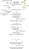
Fig. 1.
Fig. 1.
Schematic for the zero-length CX-MS data acquisition and data analysis pipeline.
Fig. 2.
Fig. 2.
Spectrin topography and minispectrin tetramer structure. (A) Schematic showing spectrin domains, the dimer–tetramer equilibria, and minispectrin. The spectrin-type domains that constitute most of the molecule are represented as rounded rectangles. Red asterisks in the minispectrin cartoon indicate the approximate location of the αL207P mutation. (B) Superimposition of the four crystal structures of spectrin-type domains [Protein Data Bank (PBD) ID: 1CUN, 1U5P, 3FB2, and 1S35] used as template building blocks for homology modeling. (C) Crystal structure for the spectrin tetramerization interface (PDB ID: 3LBX). (D) Locations of interdomain cross-links used to model minispectrin tetramer; blue lines, cross-links identified previously (21); red lines, previously unidentified cross-links; dashed lines, the same cross-links repeated in the second half of the tetramer. (E) Superimposition of present and previous tetramer structures. (F) Space-filling representations of tetramer models. β-Spectrin domains are colored in bright or pale cyan, and α-spectrin domains are colored in bright or pale orange to distinguish the two strands.
Fig. 3.
Fig. 3.
Conformational changes in the closed to open spectrin dimer transition. (A) Schematic showing the minispectrin dimer–tetramer equilibrium. (B) Dimer cross-links: (Left) specific to the closed dimer conformation; (Center) specific to the open dimer conformation; (Right) interdomain cross-links used to develop the closed dimer homology model. Dashed circles, estimated reach of the disordered N-terminal tail (yellow squiggle); red X’s, sites of cross-links to α-subunit N-terminal α-amine. (C) Dimer-specific cross-links indicative of nonhelical connectors before (Left) and after (Right) structural refinement; blue, Lys; red, Glu/Asp; green lines, cross-links with Cα–Cα distances labeled. (D) Open dimer model supported by two cross-links between α0 and α1 domains. (E) Structures showing the interconversion between fully extended open dimer to closed dimer.
Fig. 4.
Fig. 4.
Biophysical and biochemical characterization of the αL207P mutant dimer. (A) Representative sedimentation equilibrium of the WT and αL207P minispectrin. Samples were analyzed by sedimentation at 30 °C and 13,800 rpm at three different initial starting concentrations. (Upper) Residuals of the fitted curve to the actual data points from highest (black) to lowest (blue) concentration, top to bottom, respectively. (Lower) The raw data (circles) and the global fit (line) to a dimer–tetramer model. (B) HPLC gel filtration analysis of WT and αL207P minispectrin at equilibrium showing a more compact structure for αL207P dimer. (C) Representative circular dichroism data for WT and αL207P minispectrin dimers showing similar α helical content for WT and αL207P minispectrin.
Fig. 5.
Fig. 5.
Structure of the αL207P mutant dimer. (A) Locations of αL207P interdomain cross-links; asterisk, location of αL207P mutation. (B) Locations of five αL207P mutant-specific cross-links indicative of conformational rearrangements in the α1-α2-α3 region plotted on the WT structure for comparison; blue, Lys; red, Glu/Asp; black, Pro mutation; black lines, cross-links with Cα–Cα distances labeled. (C) Model of the αL207P mutant closed dimer. (D) Cα–Cα distances for interdomain cross-links identified in the αL207P mutant dimer on the WT and αL207P closed dimer structures. (E) Superimposition of α2 domains from the WT and αL207P closed dimer. The mutated residue is shown in black.

References

    1. Young MM, et al. High throughput protein fold identification by using experimental constraints derived from intramolecular cross-links and mass spectrometry. Proc Natl Acad Sci USA. 2000;97(11):5802–5806. - PMC - PubMed
    1. Chen ZA, et al. Architecture of the RNA polymerase II-TFIIF complex revealed by cross-linking and mass spectrometry. EMBO J. 2010;29(4):717–726. - PMC - PubMed
    1. Lasker K, et al. Molecular architecture of the 26S proteasome holocomplex determined by an integrative approach. Proc Natl Acad Sci USA. 2012;109(5):1380–1387. - PMC - PubMed
    1. Leitner A, et al. The molecular architecture of the eukaryotic chaperonin TRiC/CCT. Structure. 2012;20(5):814–825. - PMC - PubMed
    1. Herzog F, et al. Structural probing of a protein phosphatase 2A network by chemical cross-linking and mass spectrometry. Science. 2012;337(6100):1348–1352. - PubMed

Publication types

MeSH terms