. 2014 Feb 11;111(6):E655-62.
doi: 10.1073/pnas.1317903111. Epub 2014 Jan 22.

Structure of signaling-competent neurotensin receptor 1 obtained by directed evolution in Escherichia coli

Affiliations

Structure of signaling-competent neurotensin receptor 1 obtained by directed evolution in Escherichia coli

Pascal Egloff et al. Proc Natl Acad Sci U S A. .

Abstract

Crystallography has advanced our understanding of G protein-coupled receptors, but low expression levels and instability in solution have limited structural insights to very few selected members of this large protein family. Using neurotensin receptor 1 (NTR1) as a proof of principle, we show that two directed evolution technologies that we recently developed have the potential to overcome these problems. We purified three neurotensin-bound NTR1 variants from Escherichia coli and determined their X-ray structures at up to 2.75 Å resolution using vapor diffusion crystallization experiments. A crystallized construct was pharmacologically characterized and exhibited ligand-dependent signaling, internalization, and wild-type-like agonist and antagonist affinities. Our structures are fully consistent with all biochemically defined ligand-contacting residues, and they represent an inactive NTR1 state at the cytosolic side. They exhibit significant differences to a previously determined NTR1 structure (Protein Data Bank ID code 4GRV) in the ligand-binding pocket and by the presence of the amphipathic helix 8. A comparison of helix 8 stability determinants between NTR1 and other crystallized G protein-coupled receptors suggests that the occupancy of the canonical position of the amphipathic helix is reduced to various extents in many receptors, and we have elucidated the sequence determinants for a stable helix 8. Our analysis also provides a structural rationale for the long-known effects of C-terminal palmitoylation reactions on G protein-coupled receptor signaling, receptor maturation, and desensitization.

Keywords: detergents; membrane proteins; protein engineering; protein stability.


Conflict of interest statement

The authors declare no conflict of interest.

Figures

Fig. 1.
Fig. 1.
Structures of three evolved NTR1 variants determined devoid of fusion proteins. (A) The signaling-competent NTR1-TM86V-ΔIC3A (blue) bound to its natural agonist neurotensin (green). All selected mutations for increased expression levels in E. coli and high stability in detergent solution are depicted (orange). (B) Superposition of NTR1-TM86V-ΔIC3A (blue), NTR1-OGG7-ΔIC3A (green), and NTR1-HTGH4-ΔIC3A (orange). (C) Close-up view of the H8 region in NTR1-TM86V-ΔIC3B. Certain hydrophobic contacts of amino acids of the semiconserved H8 motif (beige) are depicted by dashed lines for clarity. The helix dipole of TM7 is illustrated by an arrow. The first of the two palmitoylation sites adjacent to the H8 C terminus is indicated. Note the absence of the palmitoyl moiety due to the prokaryotic expression. (D) Vacuum-electrostatic surface representation (PYMOL) of the neurotensin-binding pocket of TM86V-ΔIC3A. Parallel (Left) and perpendicular (Right) view to the membrane. TM5 is represented as a transparent tube in the Left panel for clarity. Neurotensin is a 13-amino-acid peptide in vivo, but only the C-terminal residues 8–13 were reported to be relevant for binding to NTR1. Strong electron density for these six amino acids was found and allowed us to model the ligand unambiguously (Fig. S2). In addition, relatively weak electron density for two N-terminal linker amino acids (Gly–Gly) of the peptide was observed in one complex of the asymmetric unit (modeled here).
Fig. 2.
Fig. 2.
Pharmacological characterizations of the crystallized NTR1 construct TM86V-ΔIC3A. (A) Neurotensin saturation-binding assay of wild-type NTR1 (circles) and TM86V-ΔIC3A (open squares). Note that Bmax levels are not representative for the expression levels of the different mutants, as 10-fold more cells were used for wild-type NTR1 to obtain a similar signal-to-noise ratio—that is, the normalized Bmax would be about 10-fold lower. (B) SR142948 antagonist competition binding experiment using wild-type NTR1 and TM86V-ΔIC3A. (C) GDP/[35S]GTPγS signaling assays of wild-type NTR1, TM86V-ΔIC3A, and TM86V-ΔIC3A L1673.50R in insect cell membranes. Equivalent amounts of active GPCR and reconstituted Gi were assayed in the presence (gray) or absence (black) of neurotensin. The signals correspond to the average of two signaling assays performed in parallel from two independent GPCR expressions, and the error bars represent SDs. (D) Pull-down experiment using immobilized Gi and solubilized GPCR from E. coli membranes. (E) Confocal imaging of living HEK293T cells expressing NTR1, TM86V-ΔIC3A, or TM86V-ΔIC3A L1673.50R-CT (reconstituted D/ERY motif and C terminus) after stimulation with fluorescent neurotensin8-13-HL647 for the indicated times.
Fig. 3.
Fig. 3.
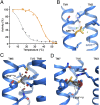
Improved interhelical surface complementarity correlates with increased thermostability. (A) Thermostability assays of NTR1-D03 (gray) and NTR1-TM86V (orange) bound to neurotensin in the harsh detergent octyl-β-d-glucopyranoside. Note that the low stability of NTR1-D03 in this detergent did not permit an accurate determination of its thermal denaturation transition point. NTR1-D03 and NTR1-TM86V are identical except for three additional mutations in NTR1-TM86V, which must confer this thermostability difference. (B–D) The structure of TM86V-ΔIC3B illustrates the 3-dimensional context at these positions. In silico back-mutating the selected residues (orange) to the wild-type amino acids (gray) would either cause a reduction of favorable van der Waals contacts (green circles in B), or it would lead to steric clashes (red circles in C and D). For the wild-type residues in C and D, the most common rotamers based on the library of PYMOL are shown. (See Fig. S8 for additional rotamers.)
Fig. 4.
Fig. 4.
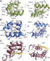
View from the cytosol onto the superposition of TM86V-ΔIC3A (blue), dark-state bovine rhodopsin (green, PDB ID code 1U19), and β2-adrenergic receptor bound to GαSβ1γ2 (salmon, PDB ID code 3SN6; GαSβ1γ2 is omitted).
Fig. 5.
Fig. 5.
Comparison of neurotensin-bound TM86V-ΔIC3B and GW5-T4L. (A) Superposition of TM86V-ΔIC3B (blue) and GW5-T4L (red), view from the intracellular side. The fused T4 lysozyme of GW5 replacing IC3 is omitted for clarity. Black arrows highlight the two different C-terminal conformations and the alternative states of TM6. (B) View along the inner leaflet of the membrane, including a part of the fused T4 lysozyme of GW5. (C and D) Comparison of the ligand-binding pockets, focusing on the interactions of EC3 with neurotensin (green). (C) The 2FO-FC omit map of TM86V-ΔIC3B (contoured at a σ level of 1.2) suggests a single α-helical turn of ECL3 in close proximity to the ligand. (D) In GW5-T4L (26) the loop contains no secondary structural element and it was modeled more distant to the peptide agonist with a cis-peptide following Asp336. Side chains of Ser335 and Gln338 were modeled up to Cβ only.
Fig. 6.
Fig. 6.
Key interactions of the H8 region in A2AR and NTR1. (A–C) Depicted are the cytosolic ends of TM1, TM2, TM7, and H8 of A2AR (A; PDB ID code 4EIY), TM86V-ΔIC3B (B), and GW5-T4L (C; PDB ID code 4GRV) viewed parallel to the membrane (Left) or from the intracellular side (Right). The yellow arrow in the GW5-T4L structure corresponds to the approximate position of H8 in TM86V-ΔIC3B.
Fig. 7.
Fig. 7.
Sequence alignment representing the end of TM7 and H8. The sequences are numbered according to Ballesteros–Weinstein (residue 8.50 chosen as F376 of NTR1). The NPxxY and F(R/K)xx(F/L)xxx(L/F) motifs are highlighted (green) and putative palmitoylation sites [experimentally confirmed in NTR1 (44, 49)] are depicted (yellow).

References

    1. Bissette G, Nemeroff CB, Loosen PT, Prange AJ, Jr, Lipton MA. Hypothermia and intolerance to cold induced by intracisternal administration of the hypothalamic peptide neurotensin. Nature. 1976;262(5569):607–609. - PubMed
    1. Schimpff RM, et al. Increased plasma neurotensin concentrations in patients with Parkinson’s disease. J Neurol Neurosurg Psychiatry. 2001;70(6):784–786. - PMC - PubMed
    1. Alifano M, et al. Neurotensin receptor 1 determines the outcome of non-small cell lung cancer. Clin Cancer Res. 2010;16(17):4401–4410. - PubMed
    1. Griebel G, Holsboer F. Neuropeptide receptor ligands as drugs for psychiatric diseases: The end of the beginning? Nat Rev Drug Discov. 2012;11(6):462–478. - PubMed
    1. Tanaka K, Masu M, Nakanishi S. Structure and functional expression of the cloned rat neurotensin receptor. Neuron. 1990;4(6):847–854. - PubMed

Publication types

Substances