. 2014 Feb 15;192(4):1361-71.
doi: 10.4049/jimmunol.1301809. Epub 2014 Jan 22.

Dectin-2 regulates the effector phase of house dust mite-elicited pulmonary inflammation independently from its role in sensitization

Affiliations

Dectin-2 regulates the effector phase of house dust mite-elicited pulmonary inflammation independently from its role in sensitization

Matthew W Parsons et al. J Immunol. .

Abstract

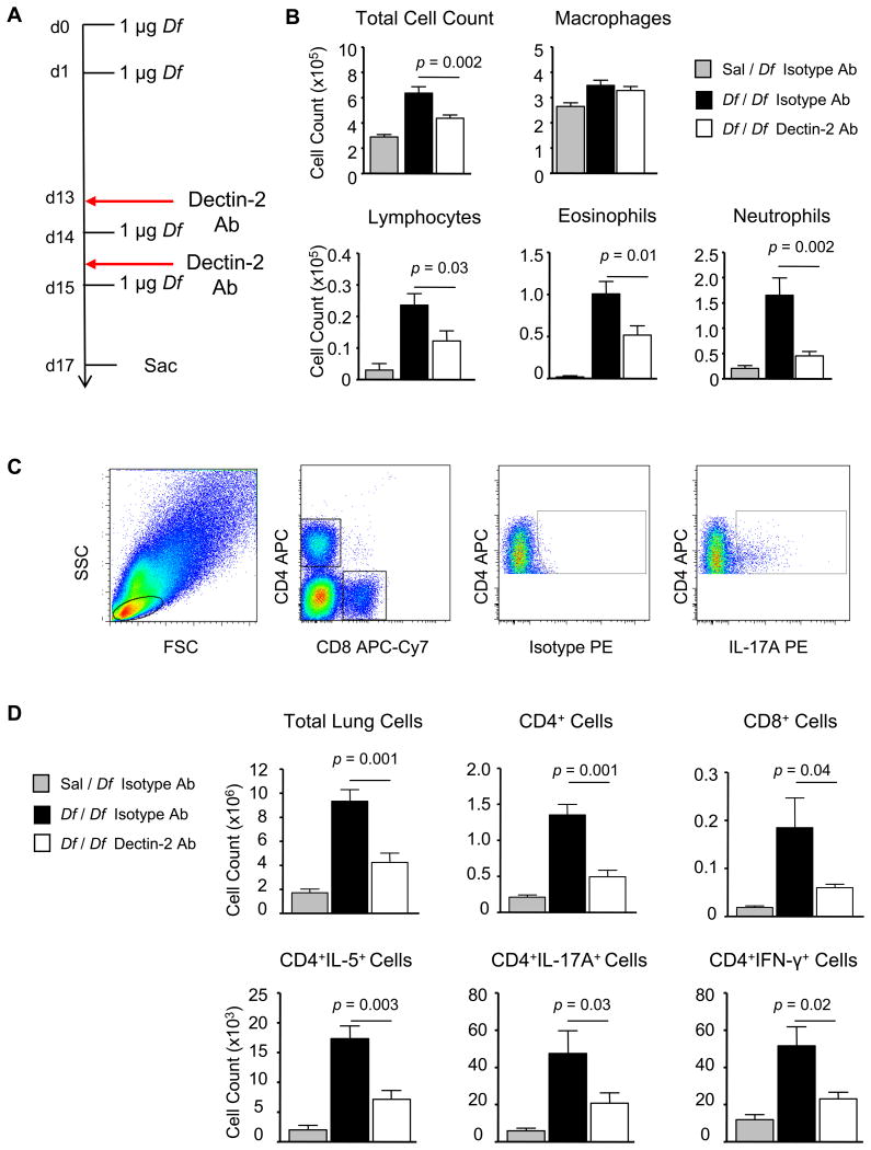
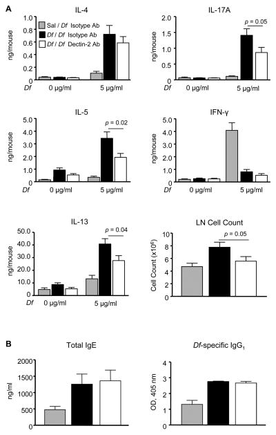
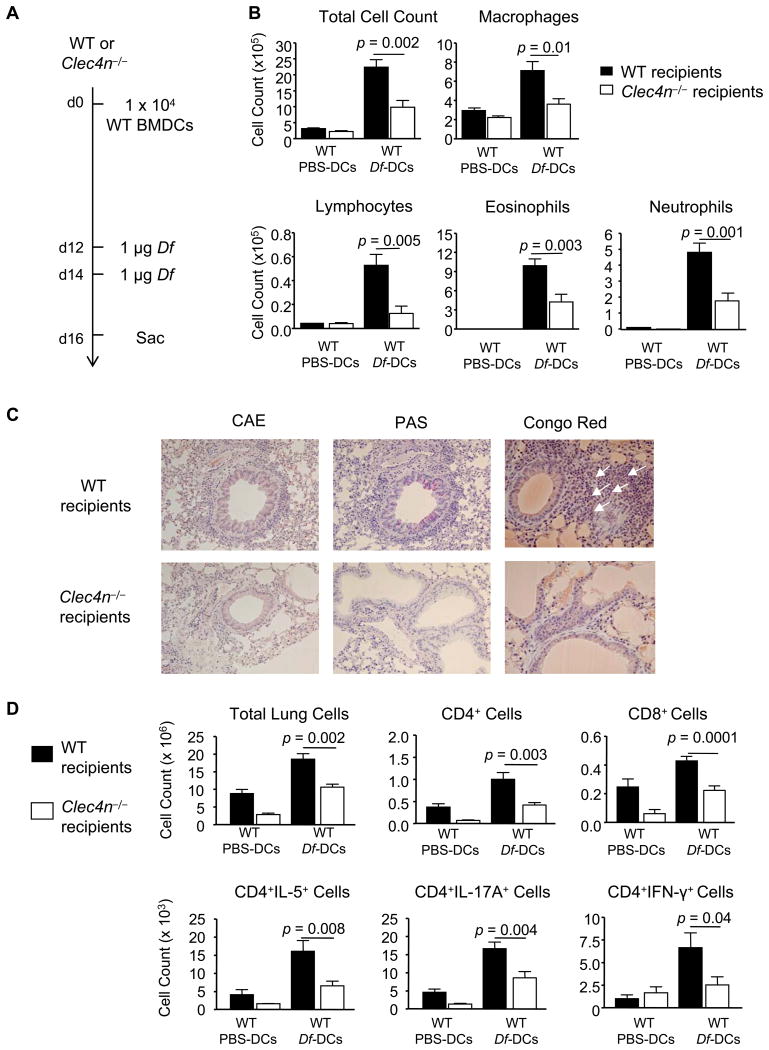
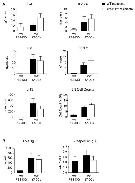
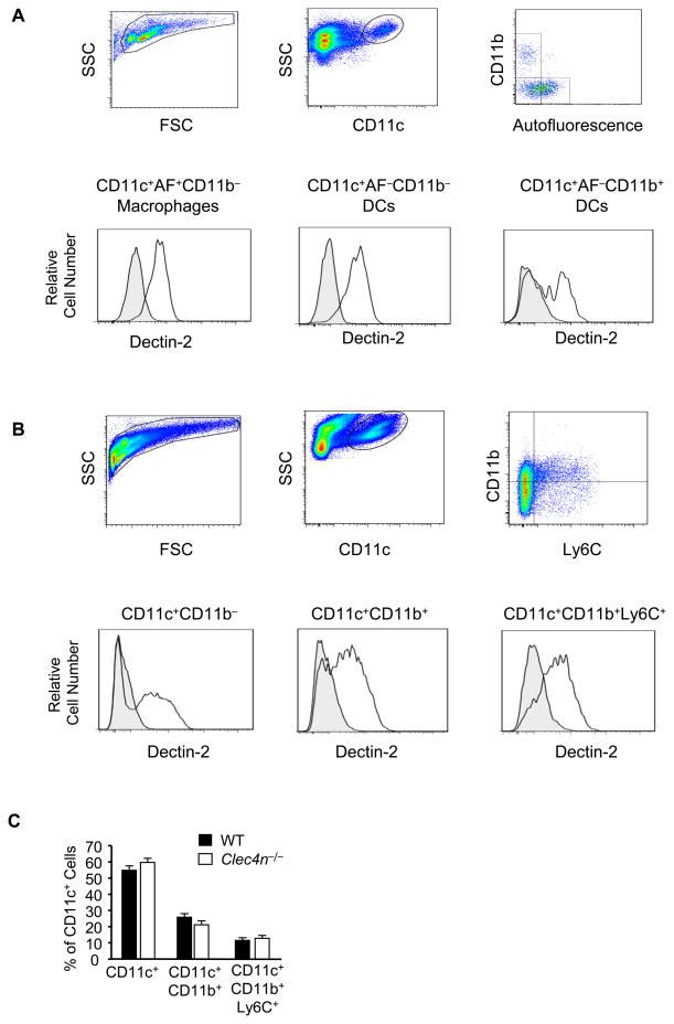
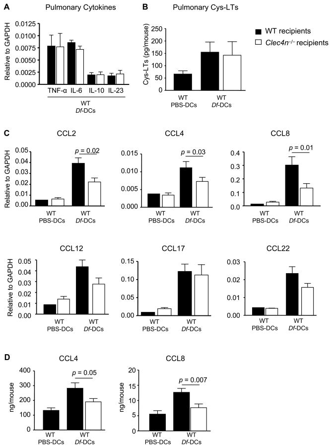
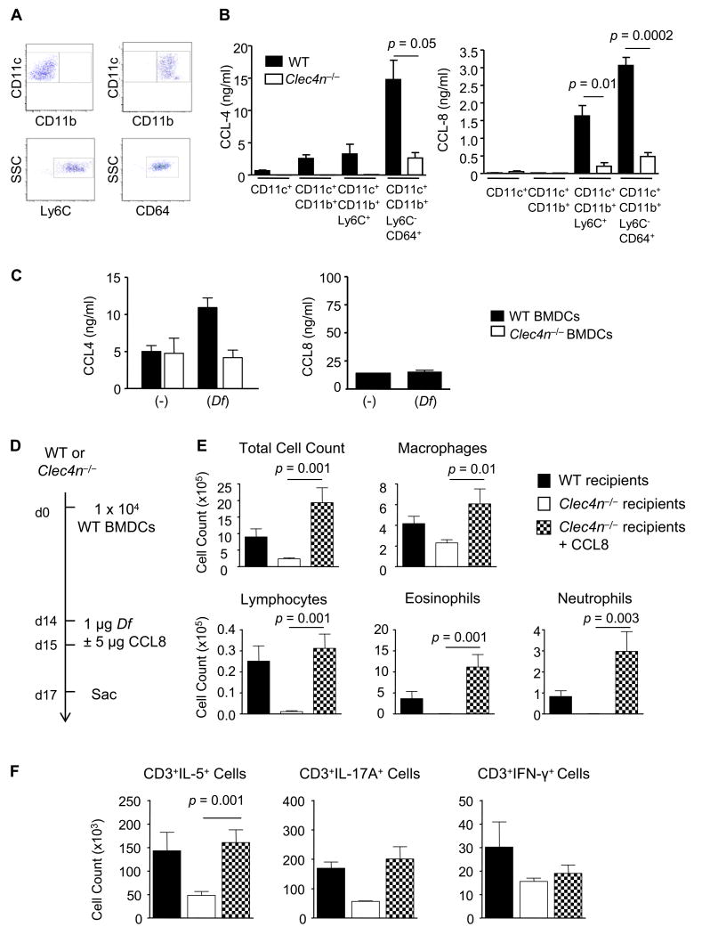
The myeloid C-type lectin receptor Dectin-2 directs the generation of Th2 and Th17 immune responses to the house dust mite Dermatophagoides farinae through the generation of cysteinyl leukotrienes and proinflammatory cytokines, respectively, but a role for Dectin-2 in effector phase responses has not been described. In this study, we demonstrate that administration of the Dectin-2 mAb solely at the time of D. farinae challenge abrogated eosinophilic and neutrophilic inflammation in the bronchoalveolar lavage fluid and Th1, Th2, and Th17 inflammation in the lung of previously sensitized mice. Furthermore, Dectin-2 null mice (Clec4n(-/-)) sensitized with the adoptive transfer of D. farinae-pulsed wild-type (WT) bone marrow-derived dendritic cells (DCs) also had less D. farinae-elicited pulmonary inflammation, supporting an effector function for Dectin-2. The protection from pulmonary inflammation seen with the Dectin-2 mAb or in Clec4n(-/-) mice was associated with little or no reduction in lung-draining lymph node cells or their cytokine production and with no reduction in serum IgE. WT and Clec4n(-/-) mice recipients, sensitized with D. farinae-pulsed WT bone marrow-derived DCs, had comparable levels of D. farinae-elicited IL-6, IL-23, TNF-α, and cysteinyl leukotrienes in the lung. By contrast, D. farinae-elicited CCL4 and CCL8 production from pulmonary CD11c(+)CD11b(+)Ly6C(+) and CD11c(+)CD11b(+)Ly6C(-)CD64(+) monocyte-derived DCs was reduced in Clec4n(-/-) recipients. Addition of CCL8 at the time of D. farinae challenge abrogated the protection from eosinophilic, neutrophilic, and Th2 pulmonary inflammation seen in Clec4n(-/-) recipients. Taken together, these results reveal that Dectin-2 regulates monocyte-derived DC function in the pulmonary microenvironment at D. farinae challenge to promote the local inflammatory response.


Figures

Figure 1
Figure 1. Dectin-2 Blocking Antibody Inhibits Df-elicited Pulmonary Inflammation in Sensitized WT Mice
WT mice were sensitized with 1 μg intranasal Df or saline control on days 0 and 1, challenged with 1 μg intranasal Df on days 14, and 15, treated with 200 μg Dectin-2 antibody or a rat IgG2a control i.p. on days 13 and 15, and killed on day 17. A. Experimental diagram. B. Cells from the BAL fluid were counted, cytospin preparations were stained, and 200 cells/slide were counted for specific cell types. Results are means ± SEM (n = 10-12 mice per group) combined from 3 independent experiments. C and D. Pulmonary mononuclear cells were isolated, stimulated with 50 ng/ml PMA and 1 μM ionomycin for 6 h in the presence of 2.5 μM monensin, stained for cell surface expression of CD4 and CD8, and for intracellular expression of IL-4, IL-5, IL-17A, and IFN-γ, and analyzed by flow cytometry. C. Representative dot plots gating on cell size, CD4 and CD8 expression, and IL-17A are shown. D. The total numbers of cells and subsets recruited to the lung are shown. Data are means ± SEM (n = 6-8 mice per group) combined from 2 independent experiments.
Figure 2
Figure 2. Dectin-2 Blocking Antibody Reduces Lymph Node Cytokine Production in Sensitized WT mice and Has No Effect on Serum Immunoglobulins
WT mice were sensitized, challenged, and treated with antibody as in Fig. 1. A. Peribronchial lymph node cells were isolated, counted, and restimulated for 72 h with 0 or 5 μg/ml Df, and cytokines in the supernatant were measured by ELISA. Data are means ± SEM (n = 10-12 mice per group) combined from 3 independent experiments. B. Serum IgE and serum Df-specific IgG1 were measured by ELISA. Data are means ± SEM (n = 10-12 mice per group) combined from 3 independent experiments.
Figure 3
Figure 3. Sensitized Clec4n−/− Mice Have Impaired Df-elicited Pulmonary Inflammation
WT or Clec4n−/− mice were sensitized with 1 × 10⁴ WT BMDC pulsed with PBS (PBS-DC) or with Df (Df-DC) and challenged with 1 μg Df on days 12 and 14. A. Experimental diagram. B. On day 16, cytospins from BAL fluid were stained and counted. Data are means ± SEM (n = 8-11/Df-DC; n = 3/PBS-DC) combined from 3 independent experiments. C. Histologic analyses of the lung. Lung tissues were fixed with paraformaldehyde and stained with the chloroacetate esterase (CAE) reaction and hematoxylin, periodic acid-Schiff (PAS), or Congo red. Eosinophils are indicated by white arrows in Congo red staining, and mucus is stained in purple in PAS. Images were acquired at 20× (CAE and PAS) and 40× (Congo red) magnifications. D. On day 16, pulmonary mononuclear cells were isolated, stimulated, fixed, and stained for CD4, CD8, and intracellular cytokines, and analyzed by flow cytometry, as in Fig. 1 C&D. Data are means ± SEM (n = 13-15/Df-DC; n = 5/PBS-DC) combined from 3 independent experiments.
Figure 4
Figure 4. Sensitized Clec4n−/− Mice Have Intact Df-Elicited Serum Immunoglobulins and Lymph Node Cytokine Production
WT or Clec4n−/− mice were sensitized and challenged as in Fig. 3. A. On day 16, peribronchial lymph node cells were isolated, counted, and restimulated for 72 h with 20 μg/ml Df, and cytokines in the supernatant were measured by ELISA. Data are means ± SEM (n = 9-11/Df-DC; n = 4/PBS-DC) combined from 3 independent experiments. B. Serum IgE and serum Df-specific IgG1 were measured by ELISA. Data are means ± SEM (n = 9-10/Df-DC; n = 4/PBS-DC) combined from 3 independent experiments.
Figure 5
Figure 5. Dectin-2 is Specifically Expressed on CD11c+ Cells in the Lung of Naive and Df-Sensitized and Challenged Mice
Single cell suspensions from the lung of WT C57BL/6 mice were generated and stained with the indicated antibodies or isotype controls. A. Top row shows gating of viable cells from naïve mice using FSC/SSC, staining for CD11c, and autofluorescence (AF)/staining for CD11b. Bottom row shows Dectin-2 expression (black lines) and isotype control (shaded gray) on CD11c+AF+CD11b macrophages (left), CD11c+AFCD11b DCs (middle) and CD11c+AFCD11b+ DCs (right). B. Top row shows gating of viable cells from WT C57BL/6 mice sensitized to 1 μg Df on day 0 and 1, challenged to 1 μg Df on days 14 and 15, and killed on day 17. FSC/SSC and stainings for CD11c, and CD11b/Ly6C are shown. Bottom row shows Dectin-2 expression (black lines) and isotype control (shaded gray) on CD11c+CD11b (left), CD11c+CD11b+ (middle), and CD11c+CD11b+Ly6C+ (right) populations. C. CD11c+ subsets from WT and Clec4n−/− lung after sensitization and challenge as in B. Data are means ± SEM (n = 4-7 mice/group) combined from 2 independent experiments.
Figure 6
Figure 6. Sensitized Clec4n−/− Mice Have Impaired Df-Elicited Pulmonary Chemokines
WT or Clec4n−/− mice were sensitized with WT BMDCs and challenged with Df, as in Figs. 3 and 4. A. Cytokine transcripts in lung 48 h after the last Df challenge. B. Cys-LTs from BAL 24 h after the last Df challenge. C. Chemokine transcripts from WT and Clec4n−/− recipients 48 h after the last Df challenge. Data are means ± SEM (n = 9-11/Df-DCs; n = 3/PBS-DCs) combined from 3 independent experiments. D. CCL4 and CCL8 from lung homogenates were measured by ELISA. Data are means ± SEM (n = 13-15/ Df-DCs; n = 4/PBS-DCs) combined from 4 independent experiments.
Figure 7
Figure 7. Sensitized Clec4n−/− Mice Have Impaired MDDC Chemokine Production and Restoration of Airway CCL8 Reconstitutes Allergic Airway Inflammation
A and B. WT or Clec4n−/− mice were sensitized with WT BMDCs and challenged with Df, as in Figs. 3 and 4. Single cell suspensions from the lung were generated, stained with the indicated antibodies or isotype controls, and sorted. A. Top row shows staining from sorted CD11c+CD11b cells (left) and CD11c+CD11b+ cells (right). Bottom row shows staining from sorted CD11c+CD11b+ Ly6C+ cells (left) and from CD11c+CD11b+ Ly6CCD64+ cells (right). B. CCL-4 (left) and CCL-8 (right) from cultured supernatant of each population at 24 h. Data are means ± SEM (n = 4-6/group) combined from 2 independent experiments. C. CCL-4 (left) and CCL-8 (right) from cultured supernatant of BMDCs with and without Df stimulation for 12 h. Data are means ± SEM (n = 4) combined from 2 independent experiments. D-F. WT or Clec4n−/− mice were sensitized with WT BMDCs and challenged with Df with or without addition of 5 μg CCL8 30 min prior to each Df challenge. D. Experimental diagram. E. Cytospins from BAL fluid were stained and counted. Data are means ± SEM (n = 7-9 mice/group) combined from 2 independent experiments. F. Pulmonary mononuclear cells were isolated, stimulated, fixed, stained for CD3 and intracellular cytokines, and analyzed by flow cytometry, as in Fig. 1 C&D. Data are means ± SEM (n = 4-7 mice/group) combined from 2 independent experiments.

Similar articles

Cited by

References

    1. Arbes SJ, Jr, Gergen PJ, Vaughn B, Zeldin DC. Asthma cases attributable to atopy: results from the Third National Health and Nutrition Examination Survey. J Allergy Clin Immunol. 2007;120:1139–1145. - PMC - PubMed
    1. Huss K, Adkinson NF, Jr, Eggleston PA, Dawson C, Van Natta ML, Hamilton RG. House dust mite and cockroach exposure are strong risk factors for positive allergy skin test responses in the Childhood Asthma Management Program. J Allergy Clin Immunol. 2001;107:48–54. - PubMed
    1. Celedon JC, Milton DK, Ramsey CD, Litonjua AA, Ryan L, Platts-Mills TAE, Gold DR. Exposure to dust mite allergen and endotoxin in early life and asthma and atopy in childhood. J Allergy Clin Immunol. 2007;120:144–149. - PMC - PubMed
    1. Hammad H, Chieppa M, Perros F, Willart MA, Germain RN, Lambrecht BN. House dust mite allergen induces asthma via Toll-like receptor 4 triggering of airway structural cells. Nat Med. 2009;15:410–416. - PMC - PubMed
    1. Trompette A, Divanovic S, Visintin A, Blanchard C, Hegde RS, Madan R, Thorne PS, Wills-Karp M, Gioannini TL, Weiss JP, Karp CL. Allergenicity resulting from functional mimicry of a Toll-like receptor complex protein. Nature. 2009;457:585–588. - PMC - PubMed

Publication types

MeSH terms