

doi: 10.1128/JVI.03264-13.
Epub 2014 Jan 22.
Evidence of in utero transmission of classical scrapie in sheep
Affiliations
- PMID: 24453368
- PMCID: PMC3993721
- DOI: 10.1128/JVI.03264-13
Item in Clipboard
Evidence of in utero transmission of classical scrapie in sheep
J Virol.
2014 Apr.
Abstract
Classical scrapie is one of the transmissible spongiform encephalopathies (TSEs), a group of fatal infectious diseases that affect the central nervous system (CNS). Classical scrapie can transmit laterally from ewe to lamb perinatally or between adult animals. Here we report detection of infectivity in tissues of an unborn fetus, providing evidence that in utero transmission of classical scrapie is also possible.
Figures
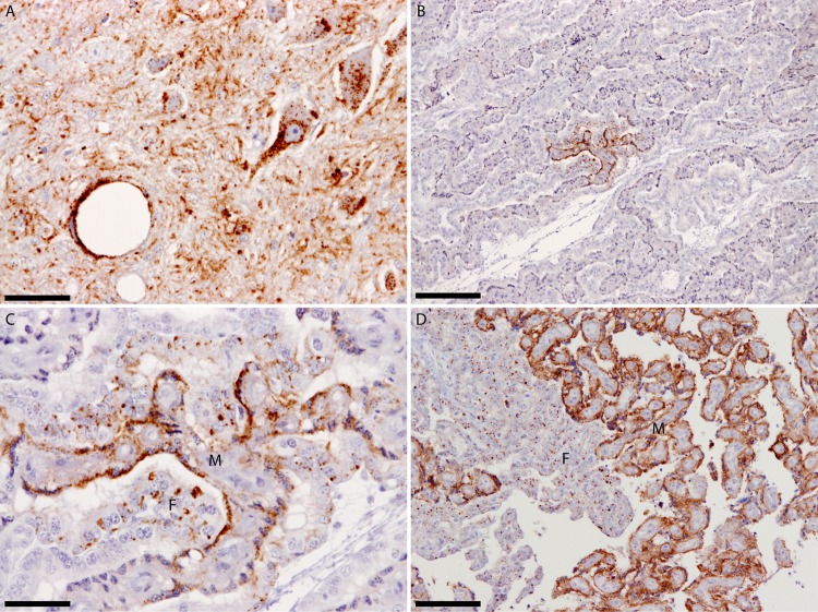
(A) Dorsal motor nucleus of the vagus nerve from ewe PG1657/05 showing widespread PrPSc deposits. (B) Placentome from fetus PG1658/05 at the 4th month of pregnancy showing restricted PrPSc deposits. (C) Greater magnification of the PrPSc-positive area shown in panel B to demonstrate that both maternal and fetal opposing units are affected and to illustrate the different PrPSc deposition patterns between maternal and fetal units. These different PrPSc deposition patterns most likely reflect the different physiological roles of maternal and fetal units, as both units were of the same PrP genotype. (D) For comparison, a placentome collected near term demonstrates the widespread PrPSc dissemination at the end of the 5th month of gestation. All sections were labeled with the monoclonal antibody R145. The maternal unit is denoted by the letter M, the fetal unit by the letter F. The scale bars in panels A and C represent 50 μm, in panels B and D, 200 μm.
Similar articles
-
Scrapie infectivity and proteinase K-resistant prion protein in sheep placenta, brain, spleen, and lymph node: implications for transmission and antemortem diagnosis.J Infect Dis. 1998 Oct;178(4):949-53. doi: 10.1086/515669. J Infect Dis. 1998. PMID: 9806020
-
PrP(Sc) detection and infectivity in semen from scrapie-infected sheep.J Gen Virol. 2012 Jun;93(Pt 6):1375-1383. doi: 10.1099/vir.0.038802-0. Epub 2012 Feb 8. J Gen Virol. 2012. PMID: 22323531
-
PrPSc accumulation in fetal cotyledons of scrapie-resistant lambs is influenced by fetus location in the uterus.J Gen Virol. 2006 Apr;87(Pt 4):1035-1041. doi: 10.1099/vir.0.81418-0. J Gen Virol. 2006. PMID: 16528055
-
Insights into Mechanisms of Transmission and Pathogenesis from Transgenic Mouse Models of Prion Diseases.Methods Mol Biol. 2017;1658:219-252. doi: 10.1007/978-1-4939-7244-9_16. Methods Mol Biol. 2017. PMID: 28861793 Free PMC article. Review.
-
Transmissible spongiform encephalopathies in food animals. Human food safety and animal feed safety concerns for veterinarians.Vet Clin North Am Food Anim Pract. 1998 Mar;14(1):49-70. doi: 10.1016/s0749-0720(15)30279-6. Vet Clin North Am Food Anim Pract. 1998. PMID: 9532667 Review.
Cited by
-
Animal prion diseases: A review of intraspecies transmission.Open Vet J. 2021 Oct-Dec;11(4):707-723. doi: 10.5455/OVJ.2021.v11.i4.23. Epub 2021 Dec 16. Open Vet J. 2021. PMID: 35070868 Free PMC article. Review.
-
Classical and Atypical Scrapie in Sheep and Goats. Review on the Etiology, Genetic Factors, Pathogenesis, Diagnosis, and Control Measures of Both Diseases.Animals (Basel). 2021 Mar 4;11(3):691. doi: 10.3390/ani11030691. Animals (Basel). 2021. PMID: 33806658 Free PMC article. Review.
-
Evaluation of Antemortem Diagnostic Techniques in Goats Naturally Infected With Scrapie.Front Vet Sci. 2020 Nov 6;7:517862. doi: 10.3389/fvets.2020.517862. eCollection 2020. Front Vet Sci. 2020. PMID: 33240943 Free PMC article.
-
How do PrPSc Prions Spread between Host Species, and within Hosts?Pathogens. 2017 Nov 24;6(4):60. doi: 10.3390/pathogens6040060. Pathogens. 2017. PMID: 29186791 Free PMC article. Review.
-
Detection of prions in oocytes and ovaries of ewes naturally infected with classical scrapie.Vet Res. 2025 Apr 10;56(1):79. doi: 10.1186/s13567-025-01512-0. Vet Res. 2025. PMID: 40211373 Free PMC article.
References
Publication types
MeSH terms
Substances
LinkOut - more resources
Full Text Sources
Other Literature Sources
Medical
