

Endobronchial perfluorocarbon reduces inflammatory activity before and after lung transplantation in an animal experimental model
- PMID: 24453412
- PMCID: PMC3888767
- DOI: 10.1155/2013/193484
Endobronchial perfluorocarbon reduces inflammatory activity before and after lung transplantation in an animal experimental model
Abstract
Background: The aim of this study was to evaluate the use of liquid perfluorocarbon (PFC) as an adjuvant substance for lung preservation and assess its role in pulmonary protection after transplantation.
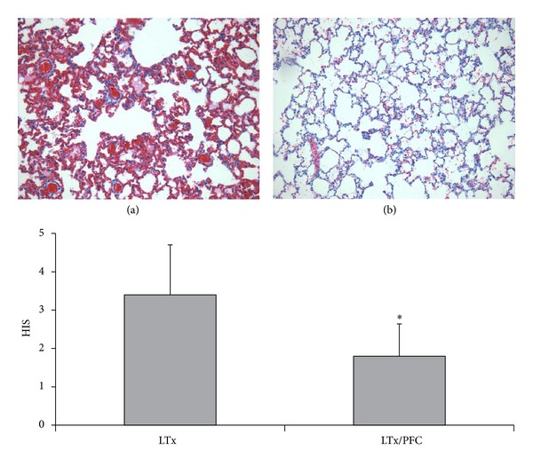
Methods: Seventy-two rat lungs were flushed with low-potassium dextran (LPD) solution and randomized into three main groups: control with LPD alone and experimental with 3 (PFC3) and 7 mL/kg (PFC7) of endobronchial PFC instilled just after harvest. Each group was divided into four subgroups according to preservation time (3, 6, 12, and 24 hours). Afterwards, we performed lung transplantation using rat lungs preserved for 12 hours with LPD alone or with 7 mL/kg of endobronchial PFC.
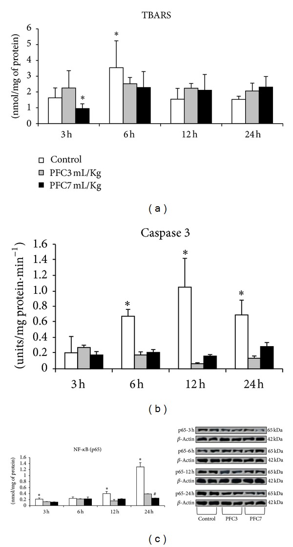
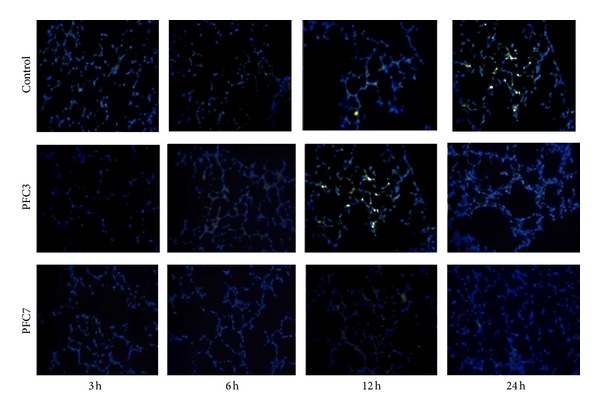
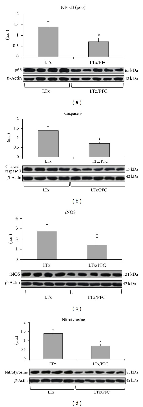
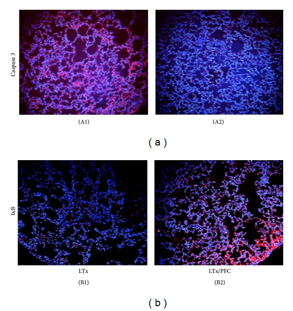
Results: There was a significant increase in oxidative stress in the control group at 6 h of cold ischemic time compared with the PFC3 and PFC7 groups. The apoptotic activity and NF-κB expression were significantly higher in the control group compared with the PFC groups at 3, 12, and 24 h of cold preservation. After transplantation, the NF-κB, iNOS, and nitrotyrosine expression as well as caspase 3 activity were significantly lower in the PFC groups.
Conclusion: The use of endobronchial PFC as an adjuvant to the current preservation strategy improved graft viability.
Figures
Similar articles
-
Effect of vaporized perfluorocarbon on oxidative stress during the cold ischemia phase of lung graft preservation.J Bras Pneumol. 2019 Mar 28;45(4):e20170288. doi: 10.1590/1806-3713/e20170288. J Bras Pneumol. 2019. PMID: 30942284 Free PMC article.
-
Endobronchial perfluorocarbon administration decreases lung injury in an experimental model of ischemia and reperfusion.J Surg Res. 2013 Aug;183(2):835-40. doi: 10.1016/j.jss.2013.01.035. Epub 2013 Feb 9. J Surg Res. 2013. PMID: 23434305
-
A new preservation solution for lung transplantation: evaluation in a porcine transplantation model.J Heart Lung Transplant. 2012 Mar;31(3):310-7. doi: 10.1016/j.healun.2011.11.009. Epub 2012 Jan 9. J Heart Lung Transplant. 2012. PMID: 22226803
-
Liver graft preservation using perfluorocarbon improves the outcomes of simulated donation after cardiac death liver transplantation in rats.Liver Transpl. 2017 Sep;23(9):1171-1185. doi: 10.1002/lt.24806. Liver Transpl. 2017. PMID: 28650112
-
Glycine intravenous donor preconditioning is superior to glycine supplementation to low-potassium dextran flush preservation and improves graft function in a large animal lung transplantation model after 24 hours of cold ischemia.J Thorac Cardiovasc Surg. 2006 Mar;131(3):724-9. doi: 10.1016/j.jtcvs.2005.09.049. J Thorac Cardiovasc Surg. 2006. PMID: 16515930
Cited by
-
Effect of vaporized perfluorocarbon on oxidative stress during the cold ischemia phase of lung graft preservation.J Bras Pneumol. 2019 Mar 28;45(4):e20170288. doi: 10.1590/1806-3713/e20170288. J Bras Pneumol. 2019. PMID: 30942284 Free PMC article.
-
Oxidative Stress and Lung Ischemia-Reperfusion Injury.Oxid Med Cell Longev. 2015;2015:590987. doi: 10.1155/2015/590987. Epub 2015 Jun 16. Oxid Med Cell Longev. 2015. PMID: 26161240 Free PMC article. Review.
-
Alpha B-crystallin Ameliorates Imbalance of Redox Homeostasis, Inflammation and Apoptosis in an Acute Lung Injury Model with Rats.Medeni Med J. 2024 Sep 30;39(3):211-220. doi: 10.4274/MMJ.galenos.2024.82274. Medeni Med J. 2024. PMID: 39350576 Free PMC article.
-
RC-3095, a selective gastrin-releasing peptide receptor antagonist, does not protect the lungs in an experimental model of lung ischemia-reperfusion injury.Biomed Res Int. 2015;2015:496378. doi: 10.1155/2015/496378. Epub 2015 Mar 29. Biomed Res Int. 2015. PMID: 25893195 Free PMC article.
References
-
- Hosenpud JD, Bennett LE, Keck BM, Boucek MM, Novick RJ. The registry of the international society for heart and lung transplantation: eighteenth official report—2001. Journal of Heart and Lung Transplantation. 2001;20(8):805–815. - PubMed
-
- Christie JD, Edwards LB, Aurora P, et al. The registry of the international society for heart and lung transplantation: twenty-sixth official adult lung and heart-lung transplantation report-2009. Journal of Heart and Lung Transplantation. 2009;28(10):1031–1049. - PubMed
-
- Sommer S-P, Sommer S, Sinha B, et al. Ischemia-reperfusion injuryinduced pulmonary mitochondrial damage. Journal of Heart and Lung Transplantation. 2011;30(7):811–818. - PubMed
-
- Cypel M, Yeung JC, Keshavjee S. Novel approaches to expanding the lung donor pool: donation after cardiac death and ex vivo conditioning. Clinics in Chest Medicine. 2011;32(2):233–244. - PubMed
Publication types
MeSH terms
Substances
LinkOut - more resources
Full Text Sources
Other Literature Sources
Medical
Research Materials
Miscellaneous
