Photoelastic analisys in the lower region of vertebral body L4
- PMID: 24453574
- PMCID: PMC3718409
- DOI: 10.1590/S1413-78522012000100003
Photoelastic analisys in the lower region of vertebral body L4
Abstract
Objective: To analyze the shear forces on the vertebral body L4 when submitted to a compression force by means of transmission photoelasticity.
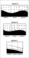
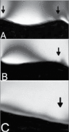
Methods: Twelve photoelastic models were divided into three groups, with four models per group, according to the positioning of the sagittal section vertebrae L4-L5 (sections A, B and C). The simulation was performed using a 15N compression force, and the fringe orders were evaluated in the vertebral body L4 by the Tardy compensation method.
Results: Photoelastic analysis showed, in general, a homogeneous distribution in the vertebral bodies. The shear forces were higher in section C than B, and higher in B than A.
Conclusion: The posterior area of L4, mainly in section C, showed higher shear concentrations, corresponding to a more susceptible area for bone fracture and spondylolisthesis. Economic and Decision Analyses - Development of an Economic or Decision Model. Level I.
Keywords: Biomechanics; Spine; Tension.
Conflict of interest statement
All the authors declare that there is no potential conflict of interest referring to this article.
Figures


References
-
- Prakash, Prabhu LV, Saralaya VV, Pai MM, Ranade AV, Singh G, et al. Vertebral body integrity: a review of various anatomical factors involved in the lumbar region. Osteoporos Int. 2007;18:891–903. - PubMed
-
- Fyhrie DP, Hoshaw SJ, Hamid MS, Hou FJ. Shear stress distribution in the trabeculae of human vertebral bone. Ann Biomed Eng. 2000;28:1194–9. - PubMed
-
- Yeni YN, Hou FJ, Vashishth D, Fyhrie DP. Trabecular shear stress in human vertebral cancellous bone: intra- and inter-individual variations. J Biomech. 2001;34:1341–6. - PubMed
-
- Lochmüller EM, Pöschl K, Würstlin L, Matsuura M, Müller R, Link TM, et al. Does thoracic or lumbar spine bone architecture predict vertebral failure strength more accurately than density? Osteoporos Int. 2008;19:537–45. - PubMed
LinkOut - more resources
Full Text Sources
