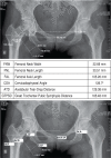
Radiographic anatomy of the proximal femur: correlation with the occurrence of fractures
- PMID: 24453585
- PMCID: PMC3718425
- DOI: 10.1590/S1413-78522012000200004
Radiographic anatomy of the proximal femur: correlation with the occurrence of fractures
Abstract
Objective: To evaluate the correlation between radiographic parameters of the proximal femur anatomy and fractures.
Methods: Three hundred and five digital x-rays of the pelvis were analyzed in the anteroposterior view. Of these x-rays, twenty-seven showed femoral neck or transtrochanteric fractures. The anatomical parameters analyzed were: femoral neck width (FNW), femoral neck length (FNL), femoral axis length (FAL), cervicodiaphyseal angle (CDA), acetabular tear-drop distance (ATD) and great trochanter-pubic symphysis distance (GTPSD). The analysis was performed by comparing the results of the x-rays with and without proximal femoral fracture, to establish a correlation between them.
Results: No differences were found between the anatomical parameters of the groups with and without proximal femoral fracture.
Conclusion: There was no association between anatomical changes in the proximal femur and greater susceptibility to fractures. Level of evidence IV, Cross-sectional Study.
Keywords: Femoral fractures; Femur; Femur neck; Radiography.
Conflict of interest statement
All the authors declare that there is no potential conflict of interest referring to this article.
Figures
References
-
- Baumgaertner MR, Higgins TF.Fratura do colo do fêmur In: Rockwood CA Jr, Green DP, Bucholz RW.Rockwood e Green fraturas em adultos. 5th ed. Philadelphia: JB Lippincott; 2006. p.1579-8
-
- Koval KJ, Zuckerman JD.Fraturas intertrocantéricas In: Rockwood CA Jr, Green DP, Bucholz RW.Rockwood e Green fraturas em adultos. 5th ed. Philadelphia: JB Lippincott; 2006. 1635-80
-
- Mourão AL, Vasconcellos HA. Geometria do fêmur proximal em ossos de brasileiros. Acta Fisiátrica. 2001;8(3):113–8.
-
- Canto RS, Silveira MA, Rosa AS, Gomide LC, Baraúna MA. Morfologia radiográfica de quadril e pelve e sua relação com fraturas proximais do fêmur. Rev Bras Ortop. 2003;38(1/2):12–20.
-
- Gnudi S, Ripamonti C, Gualtieri G, Malavolta N. Geometry of proximal femur in the prediction of hip fracture in osteoporotic women. Br J Radiol. 1999;72(860):729–33. - PubMed
LinkOut - more resources
Full Text Sources
Research Materials

