

Study of the anatomy of the tibial nerve and its branches in the distal medial leg
- PMID: 24453596
- PMCID: PMC3718430
- DOI: 10.1590/S1413-78522012000300005
Study of the anatomy of the tibial nerve and its branches in the distal medial leg
Abstract
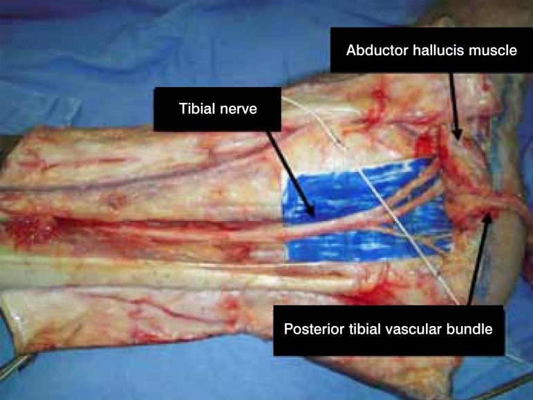
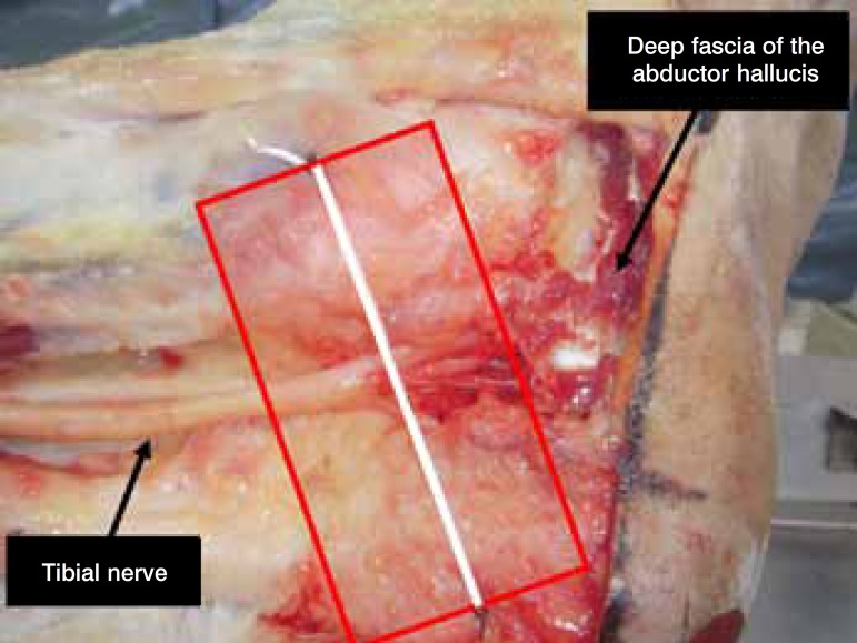
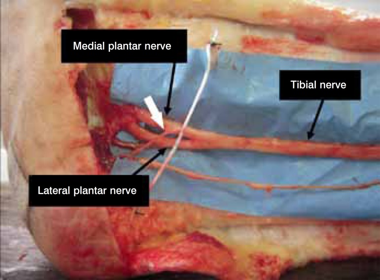
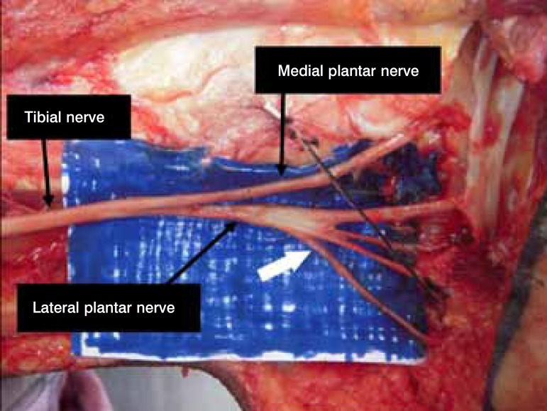
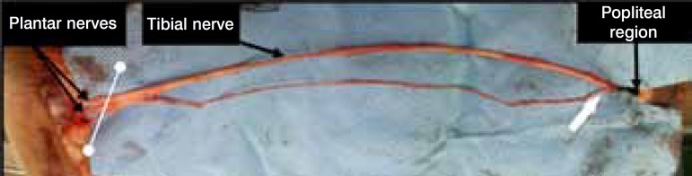
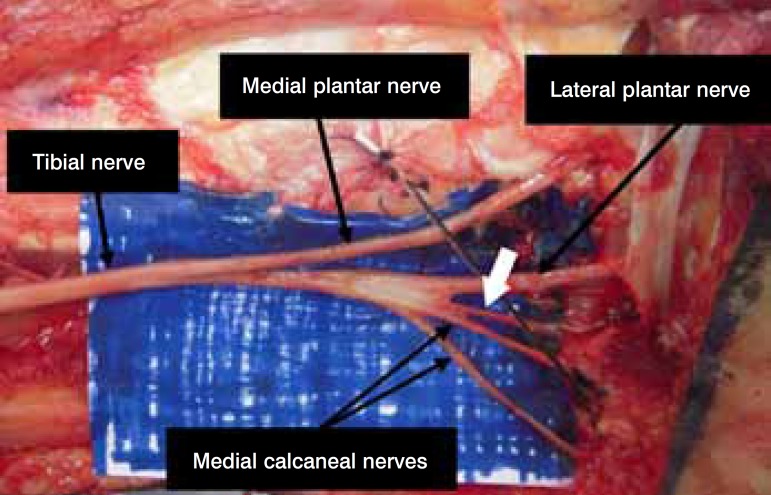
Objective: Determine, through dissection in fresh cadavers, the topographic anatomy of the tibial nerve and its branches at the ankle, in relation to the tarsal tunnel.
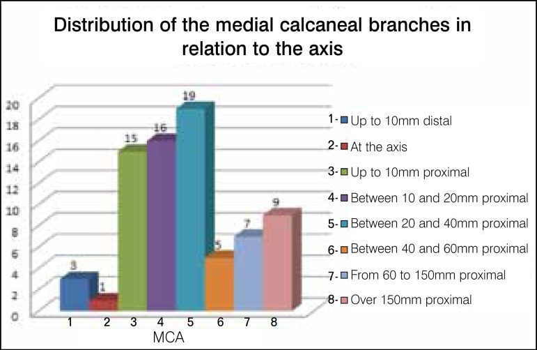
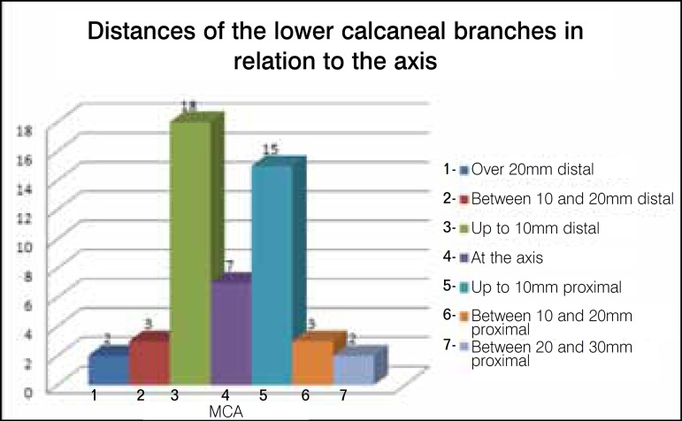
Methods: Bilateral dissections were performed on 26 fresh cadavers and the locations of the tibial nerve bifurcation and its branches were measured in millimeters. For the calcaneal branches, the amount and their respective nerves of origin were also analyzed.
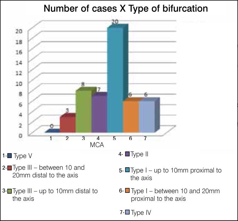
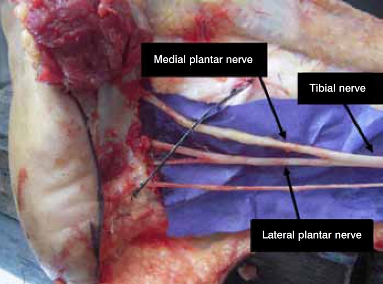
Results: The tibial nerve bifurcation occurred under the tunnel in 88% of the cases and proximally in 12%. As for the calcaneal branches, the medial presented with one (58%), two (34%) and three (8%) branches, with the most common source occurring in the tibial nerve (90%) and the lower with a single branch per leg and lateral plantar nerve as the most common origin (70%). Level of Evidence, V Expert opinion .
Keywords: Cadáver; Dissection; Peripheral nerves; Tarsal tunnel syndrome; Tibial nerve/anatomy & histology.
Conflict of interest statement
All the authors declare that there is no potential conflict of interest referring to this article.
Figures
References
-
- Kopell HP, Thompson WA. [Peripheral entrapment neuropathies of the lower extremity.] N Engl J Med. 1960;262:56–60. - PubMed
-
- Keck C. The Tarsal-Tunnel syndrome. Bone Joint Surgery. 1962;44:180–2.
-
- Lam SJ. A tarsal-tunnel syndrome. Lancet. 1962;2(7270):1354–5. - PubMed
-
- Upton AR, McComas AJ. The double crush in nerve entrapment syndromes. Lancet. 1973;2(7825):359–62. - PubMed
-
- Dellon AL, Mackinnon SE. Chronic nerve compression model for the double crush hypothesis. Ann Plast Surg. 1991;26(3):259–64. - PubMed
LinkOut - more resources
Full Text Sources
Research Materials