

Extracellular matrix remodeling in experimental intervertebral disc degeneration
- PMID: 24453658
- PMCID: PMC3861996
- DOI: 10.1590/S1413-78522013000300003
Extracellular matrix remodeling in experimental intervertebral disc degeneration
Abstract
Objective: To evaluate the remodeling of the extracellular matrix in intervertebral disc degeneration through the experimental model of intervertebral disc degeneration.
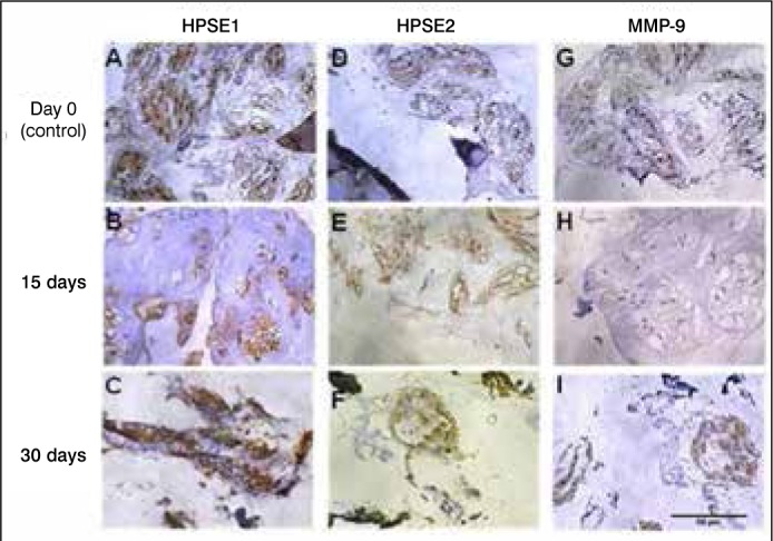
Methods: The model of disc degeneration induction, using needle 20G and 360° rotation, was applied for 30 seconds between the 6(th)/7(th), and 8(th)/9(th) coccygeal vertebrae of Wistar rats. The intermediary level, between the 7(th) and 8(th) vertebrae, was taken as control, not being subjected puncture. The distribution of the extracellular matrix components involved in the remodeling and inflammation process, such as proteoglycans (aggrecan, decorin, biglycan), growth factors (TGFβ), heparanase isoforms (HPSE1, HPSE2), metaloprotesasis-9 (MMP9) and interleukins (IL-6, IL-10) was analyzed during the post-injury period (15 to 30 days) and in the control group (discs collected immediately after the puncture, day zero). On the 15(th) day, acute phase of the disease, a reduced expression of extracellular matrix components had been observed, whilst there were no differences in the interleukins expression. At 30 days, the molecules followed a very similar pattern of expression in the control group (not affected by disc degeneration).
Results: The results show that during the acute phase significant alterations in the extracellular matrix components occur and in the late phase intervertebral disc returns to a profile similar to noninvolved tissue, probably due to extensive remodeling process of the extracellular matrix that is capable of regenerating the damaged tissue.
Conclusion: : The experimental model used demonstrated the occurrence of significant changes in the extracellular matrix during the period analyzed after induction of intervertebral disc degeneration. Laboratory investigation.
Keywords: Interleukins; Intervertebral disc degeneration; Proteoglycans; Rats, Wistar.
Conflict of interest statement
All the authors declare that there is no potential conflict of interest referring to this article.
Figures


Similar articles
-
STUDIES OF MOLECULAR CHANGES IN INTERVERTEBRAL DISC DEGENERATION IN ANIMAL MODEL.Acta Ortop Bras. 2016 Jan-Feb;24(1):16-21. doi: 10.1590/1413-785220162401152960. Acta Ortop Bras. 2016. PMID: 26997908 Free PMC article.
-
Expression of heparanase isoforms in intervertebral discs classified according to Pfirrmann grading system for disc degeneration.Spine (Phila Pa 1976). 2013 Jun 1;38(13):1112-8. doi: 10.1097/BRS.0b013e3182894cf4. Spine (Phila Pa 1976). 2013. PMID: 23370684
-
Changes in mRNA and protein levels of proteoglycans of the anulus fibrosus and nucleus pulposus during intervertebral disc degeneration.Spine (Phila Pa 1976). 2002 Oct 15;27(20):2212-9. doi: 10.1097/00007632-200210150-00006. Spine (Phila Pa 1976). 2002. PMID: 12394896
-
Roles of large aggregating proteoglycans in human intervertebral disc degeneration.Connect Tissue Res. 2019 May;60(3):209-218. doi: 10.1080/03008207.2018.1499731. Epub 2018 Aug 9. Connect Tissue Res. 2019. PMID: 29992840 Review.
-
The aging spine: the role of inflammatory mediators in intervertebral disc degeneration.Cell Mol Biol (Noisy-le-grand). 2007 May 30;53(5):4-18. Cell Mol Biol (Noisy-le-grand). 2007. PMID: 17543240 Review.
Cited by
-
Protective effect of ligustrazine on lumbar intervertebral disc degeneration of rats induced by prolonged upright posture.Evid Based Complement Alternat Med. 2014;2014:508461. doi: 10.1155/2014/508461. Epub 2014 Apr 29. Evid Based Complement Alternat Med. 2014. PMID: 24872832 Free PMC article.
-
STUDIES OF MOLECULAR CHANGES IN INTERVERTEBRAL DISC DEGENERATION IN ANIMAL MODEL.Acta Ortop Bras. 2016 Jan-Feb;24(1):16-21. doi: 10.1590/1413-785220162401152960. Acta Ortop Bras. 2016. PMID: 26997908 Free PMC article.
-
Extra Cellular Matrix Remodeling: An Adjunctive Target for Spinal Cord Injury and Intervertebral Disc Degeneration.Neurospine. 2022 Sep;19(3):632-645. doi: 10.14245/ns.2244366.183. Epub 2022 Sep 30. Neurospine. 2022. PMID: 36203290 Free PMC article.
-
Altered serum cytokines in patients with symptomatic disk herniation and depressive symptoms.Front Neurosci. 2024 Apr 5;18:1366559. doi: 10.3389/fnins.2024.1366559. eCollection 2024. Front Neurosci. 2024. PMID: 38646609 Free PMC article.
-
Comprehensive analysis of potential ceRNA network and immune cell infiltration in intervertebral disc degeneration.J Orthop Surg Res. 2022 Sep 29;17(1):432. doi: 10.1186/s13018-022-03331-x. J Orthop Surg Res. 2022. PMID: 36175893 Free PMC article.
References
-
- Rodrigues LM, Rachel TT, Mader AM, Milani C, Ueno FH, Pinhal MAS. Análise comparativa Histopatológica entre hernia de disco contida e extrusa. Coluna/Columna. 2011; 10 (1):55–57.
-
- Cs-Szabo G, Ragasa-San Juan D, Turumella V, Masuda K, Thonar EJ, An HS. Changes in mRNA and protein levels of proteoglycans of the anulus fibrosus and nucleus pulposus during intervertebral disc degeneration. Spine (Phila Pa 1976) 2002;27(20):2212–2219. - PubMed
-
- Pearce RH, Grimmer BJ, Adams ME. Degeneration and the chemical composition of the human lumbar intervertebral disc. J Orthop Res. 1987;5(2):198–205. - PubMed
LinkOut - more resources
Full Text Sources
Other Literature Sources
Miscellaneous