. 2014 Jan;10(1):e1003435.
doi: 10.1371/journal.pcbi.1003435. Epub 2014 Jan 16.

Coarse-grained molecular simulation of epidermal growth factor receptor protein tyrosine kinase multi-site self-phosphorylation

Affiliations

Coarse-grained molecular simulation of epidermal growth factor receptor protein tyrosine kinase multi-site self-phosphorylation

John G Koland. PLoS Comput Biol. 2014 Jan.

Abstract

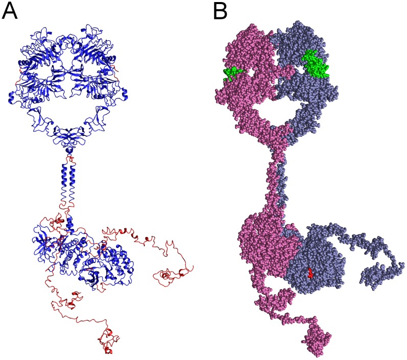
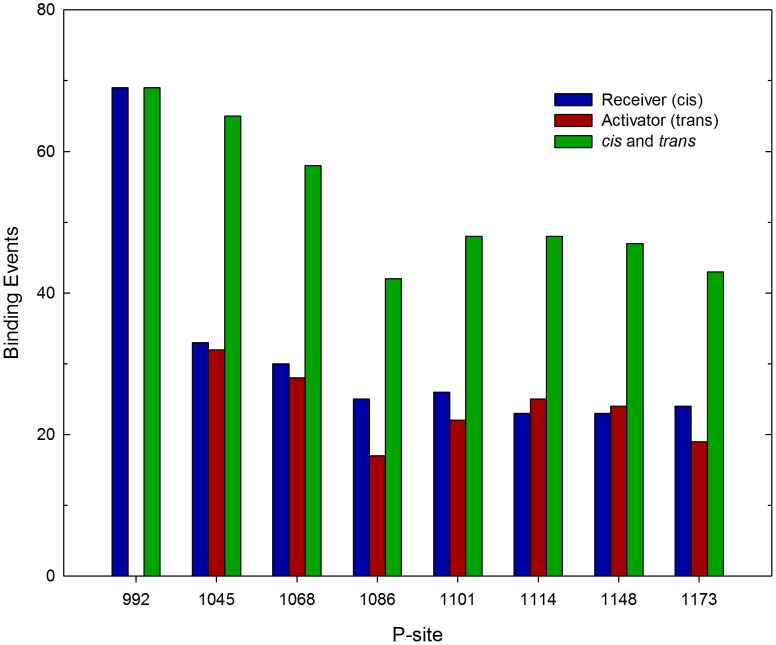
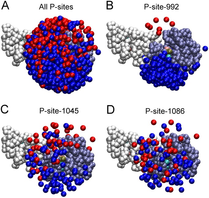
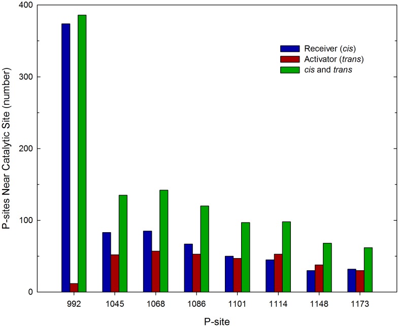
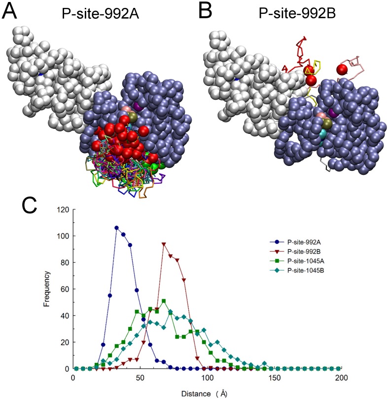
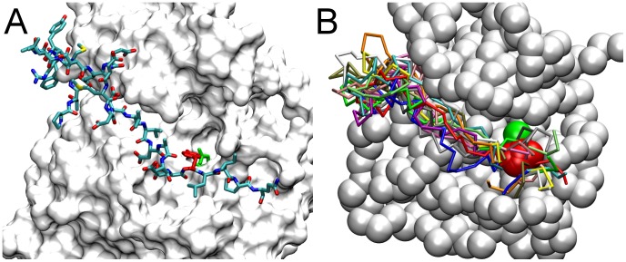
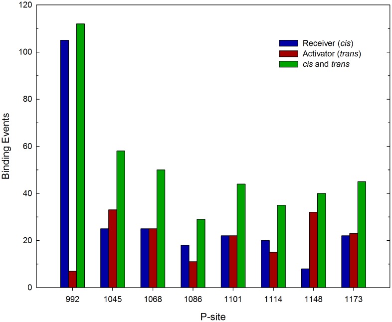
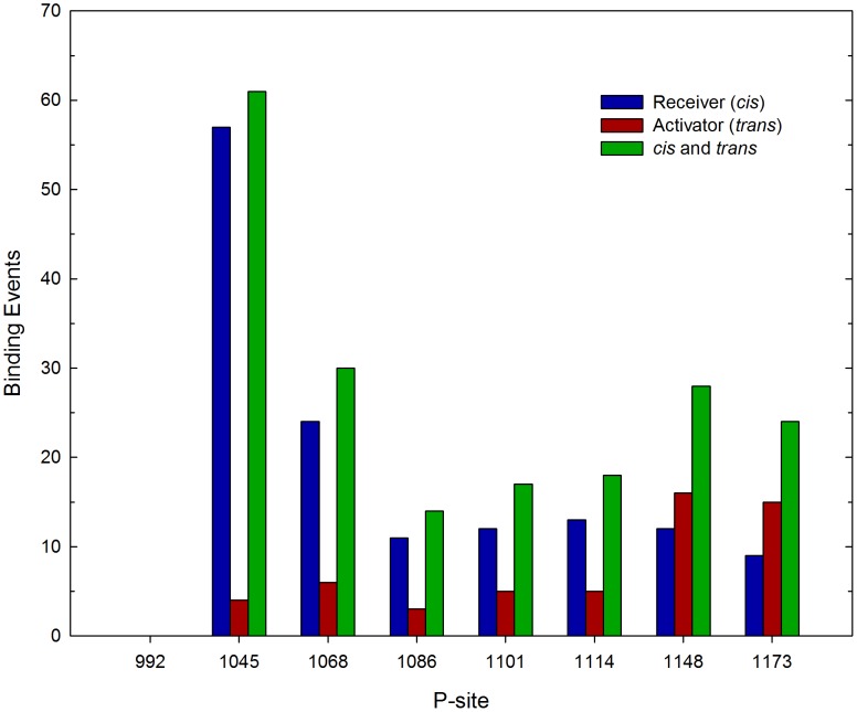
Upon the ligand-dependent dimerization of the epidermal growth factor receptor (EGFR), the intrinsic protein tyrosine kinase (PTK) activity of one receptor monomer is activated, and the dimeric receptor undergoes self-phosphorylation at any of eight candidate phosphorylation sites (P-sites) in either of the two C-terminal (CT) domains. While the structures of the extracellular ligand binding and intracellular PTK domains are known, that of the ∼225-amino acid CT domain is not, presumably because it is disordered. Receptor phosphorylation on CT domain P-sites is critical in signaling because of the binding of specific signaling effector molecules to individual phosphorylated P-sites. To investigate how the combination of conventional substrate recognition and the unique topological factors involved in the CT domain self-phosphorylation reaction lead to selectivity in P-site phosphorylation, we performed coarse-grained molecular simulations of the P-site/catalytic site binding reactions that precede EGFR self-phosphorylation events. Our results indicate that self-phosphorylation of the dimeric EGFR, although generally believed to occur in trans, may well occur with a similar efficiency in cis, with the P-sites of both receptor monomers being phosphorylated to a similar extent. An exception was the case of the most kinase-proximal P-site-992, the catalytic site binding of which occurred exclusively in cis via an intramolecular reaction. We discovered that the in cis interaction of P-site-992 with the catalytic site was facilitated by a cleft between the N-terminal and C-terminal lobes of the PTK domain that allows the short CT domain sequence tethering P-site-992 to the PTK core to reach the catalytic site. Our work provides several new mechanistic insights into the EGFR self-phosphorylation reaction, and demonstrates the potential of coarse-grained molecular simulation approaches for investigating the complexities of self-phosphorylation in molecules such as EGFR (HER/ErbB) family receptors and growth factor receptor PTKs in general.


Conflict of interest statement

The author has declared that no competing interests exist.

Figures

Figure 1
Figure 1. Complete structural model of the dimeric EGFR.
Shown in two representations is the complete structural model of a dimer of EGFR molecules, each with a bound EGF molecule and bound AMPPNP.2Mg2+ substrate complex, generated herein. (A) Backbone conformation and secondary structure of the modeled EGFR polypeptides, with segments derived from published crystallographic structures colored blue and modeled segments colored red. (B) Van der Waals representation of the EGFR dimer, with the monomers having active (receiver) and inactive (activator) conformation kinases colored ice-blue and mauve, respectively, bound EGF molecules colored green, and bound AMPPNP.2Mg2+ substrate complexes colored red.
Figure 2
Figure 2. Active conformation PTK domain with bound nucleotide and peptide substrates in the complete EGFR model.
Shown in an accessible surface representation are residues of the active conformation PTK domain (white) with its catalytic Asp-813 and bound AMPPNP.2Mg2+ substrate complex highlighted in CPK coloring. The docked nine-amino acid P-site-1173 peptide (sequence ENAEYLRVA) is shown in green. Note that the tyrosine hydroxyl of the peptide substrate is in close proximity to both the carboxyl group of Asp-813 and the γ-phosphate of the AMPPNP substrate analog. Similar models were generated with each of the eight distinct P-site peptides (see Table 1) docked in the active site.
Figure 3
Figure 3. Conformation of the CT domains in randomized dimeric EGFR structural models.
(A) Rendering of fifty randomized structures of the coarse-grained EGFR model showing the backbone conformation of the intracellular (in a different color for each structure) and TM (all in yellow) domains, with the TM domains of each structure together aligned. (B) Graphic of the same structures with the active conformation kinase domain of the receiver molecule in each aligned, the CT domains of the receiver (residues 968 to 1186) and activator (residues 960 to 1186) molecules colored in blue and red, respectively, and pseudo-atoms of the receiver (residues 679 to 967) and activator (residues 679 to 959) kinase domains depicted as ice-blue and white beads, respectively.
Figure 4
Figure 4. Frequency of P-site/catalytic site interactions in iterative simulations of the EGFR self-phosphorylation event.
The frequencies of stable interactions of individual P-sites with the catalytic site were tabulated following 420 total molecular simulations, each performed using an initial structure randomly selected from a set of five hundred dimeric EGFR structures with randomized CT domain conformations (see Fig. 3). Interactions of individual P-sites in the CT domains of the receiver and activator molecules are separately tabulated, as are the sums of interactions of the P-sites in both chains (cis and trans). Note there was a general trend of reduced frequency of P-site/catalytic site binding events with increasing residue number, with the kinase-proximal site P-site-992 showing the most frequent interactions. Also, the catalytic site interactions of P-site-992 occurred exclusively in cis versus in trans.
Figure 5
Figure 5. Proximity of P-site tyrosine residues to the catalytic site in randomized EGFR structural models.
Renderings of the five hundred randomized structures used as initial structures in simulations after alignment of their receiver kinase domains are shown. Pseudo-atoms of one receiver (residues 679 to 967) and one activator (residues 679 to 959) kinase domain are depicted as ice-blue and white beads, respectively, and those representing P-site tyrosines in the CT domains of receiver and activator molecules depicted as blue and red beads, respectively. (A) Shown are those pseudo-atoms of all P-site tyrosine residues within 40 Å of the γ-phosphate of the AMPPMP substrate (tan bead) bound in the catalytic site of the receiver molecule. (B–D) Similar renderings but with only the tyrosines of P-site-992 (B), -1045 (C) or -1086 (D) shown. Note an apparent bias in the access of P-site-922 of the receiver molecule to the catalytic site. The number of P-sites of each variety in the vicinity of the catalytic site is quantified in Fig. 6.
Figure 6
Figure 6. Number of P-sites in proximity of the catalytic site in randomized EGFR structural models.
The set of five hundred dimeric EGFR structures with randomized CT domain conformations was analyzed to determine the number of structures that had a given P-site tyrosine residue within 40 Å of the γ-phosphate of the AMPPMP substrate bound in the catalytic site. The kinase-proximal site P-site-992 of the receiver molecule was most often and that of the activator molecule least often near the catalytic site, consistent with the bias for cis versus trans P-site-992 binding events in simulations (cf. Fig. 4). This cis-trans bias, as well as the number in proximity of the catalytic site, was markedly reduced for P-sites more distal to the kinase core.
Figure 7
Figure 7. Relative catalytic site access of P-site-992 of the receiver and activator molecules.
Structures were examined among a set of 650 selected at 100 µsec molecular trajectories of EGFR dimer motion (from simulations in which P-site/catalytic site residues pairs were not considered to form native contacts), to identify subsets in which P-site Tyr-992 of the receiver and activator molecules were within 30 Å of catalytic Asp-813 of the receiver. Compared here are the identified catalytic site excursions of P-site-922 of the receiver (A) and activator (B) molecules, after alignment of receiver PTK domains (residues 679 to 967) in the identified structures. One receiver and one activator PTK domain are shown as ice-blue and white beads, respectively, with the catalytic Asp-813 as a purple bead and the bound nucleotide substrate complex as beads with CPK coloring. The backbones of the partial CT domains of receiver (residues 968 to 991) and activator (residues 960 to 991) molecules are shown in different colors, with residue 967 or 959 at the origin of these domains and their target Tyr-992 depicted as green and red beads, respectively. Comparison of the two panels indicates that P-site-992 of the receiver made much more frequent excursions near the catalytic site. (C) Distances from the pseudo-atoms representing tyrosine residues of the indicated P-sites to the γ-phosphate of the AMPPMP substrate bound in the catalytic site were quantified for each of the five hundred randomized structural models used as initial structures for simulations and the distance distributions plotted. Note that while there was an obvious bias in the catalytic site proximities of the receiver P-site-992 (P-site-992A) versus that of the activator (P-site-992B), this bias was markedly reduced for P-sites more distal to the kinase core (e.g. P-site-1045A versus -1045B).
Figure 8
Figure 8. A substrate binding cleft facilitates the cis interaction of the kinase-proximal P-site-992 with the catalytic site.
(A) After docking the P-site-992 peptide (residues 988 to 996) in the catalytic site, the structure of the CT domain sequence between it and the kinase core (residues 968 to 987) was modeled with the program Loopy (see Materials and Methods). The resulting all-atom structure is shown with the solvent accessible surface of PTK domain residues 669 to 967 colored white, modeled CT domain residues 968 to 996 in CPK coloring, and the target Tyr-992 and catalytic Asp-813 residues highlighted in red and green, respectively. The modeled CT domain sequence was found to thread through a cleft between the N- and C-terminal lobes of the PTK domain, suggesting that the presence of this cleft facilitates access in cis of the kinase-proximal P-site-992 to the catalytic site. (B) In simulations of P-site/catalytic site binding events (Fig. 4), the most frequent events (69 of 420) were cis interactions of P-site-992 of the receiver molecule with the catalytic site. Shown here are the final structures from fourteen simulations randomly chosen from those ending with a cis P-site-992 binding event, after alignment of the receiver PTK domain pseudo-atoms 679 to 967 (those of one PTK domain shown as chalk-white beads). The backbone conformations of each of the fourteen CT domain sequences (residues 968 to 996) are shown in different colors, with the target Tyr-992 and catalytic Asp-813 depicted as red and green beads, respectively.
Figure 9
Figure 9. Effect of CT domain electrostatic interactions on the frequency of P-site/catalytic site binding events.
The frequency of stable interactions of individual P-sites with the catalytic site was tabulated following 413 total simulations performed with a model in which all CT domain charges were eliminated. Interactions with the catalytic site of individual P-sites in the CT domains of the receiver and activator molecules are separately tabulated, as are the sum of interactions of the P-sites in both chains (cis and trans). Compared to what was seen in simulations with CT domain charges included (cf. Fig. 4), P-site-922 had a much enhanced tendency for in cis catalytic site interactions.
Figure 10
Figure 10. Second P-site binding event following an initial binding of P-site-992A.
Repeated simulations were initiated with structures representing the dimeric EGFR with P-site-992A of the receiver monomer bound in the catalytic site (see Fig. 8B), and with the nascent phosphorylation of P-site-992A mimicked by the introduction of a negative charge and the removal of its P-site/active site interaction potentials in the simulation model (see text). Shown here are the frequencies with which individual P-sites underwent a subsequent P-site binding event over a course of 192 total simulations. A notable bias in favor of cis (Receiver, n = 138) versus trans (Activator, n = 54) binding events was seen, with the sites closest in sequence to P-site-992 of the receiver molecule binding most frequently.

Similar articles

Cited by

References

    1. Hubbard SR, Miller WT (2007) Receptor tyrosine kinases: mechanisms of activation and signaling. Curr Opin Cell Biol 19: 117–123 - PMC - PubMed
    1. Ferguson KM (2008) Structure-based view of epidermal growth factor receptor regulation. Annu Rev Biophys 37: 353–373 - PMC - PubMed
    1. Jura N, Endres NF, Engel K, Deindl S, Das R, et al. (2009) Mechanism for activation of the EGF receptor catalytic domain by the juxtamembrane segment. Cell 137: 1293–1307 - PMC - PubMed
    1. Zhang X, Gureasko J, Shen K, Cole PA, Kuriyan J (2006) An allosteric mechanism for activation of the kinase domain of epidermal growth factor receptor. Cell 125: 1137–1149 - PubMed
    1. Margolis BL, Lax I, Kris R, Dombalagian M, Honegger AM, et al. (1989) All autophosphorylation sites of epidermal growth factor receptor and HER2/neu are located in their carboxyl-terminal tails: identification of a novel site in EGF receptor. J Biol Chem 264: 10667–10671 - PubMed

Publication types