. 2014 Jan;10(1):e1003847.
doi: 10.1371/journal.ppat.1003847. Epub 2014 Jan 16.

KSHV 2.0: a comprehensive annotation of the Kaposi's sarcoma-associated herpesvirus genome using next-generation sequencing reveals novel genomic and functional features

Affiliations

KSHV 2.0: a comprehensive annotation of the Kaposi's sarcoma-associated herpesvirus genome using next-generation sequencing reveals novel genomic and functional features

Carolina Arias et al. PLoS Pathog. 2014 Jan.

Abstract

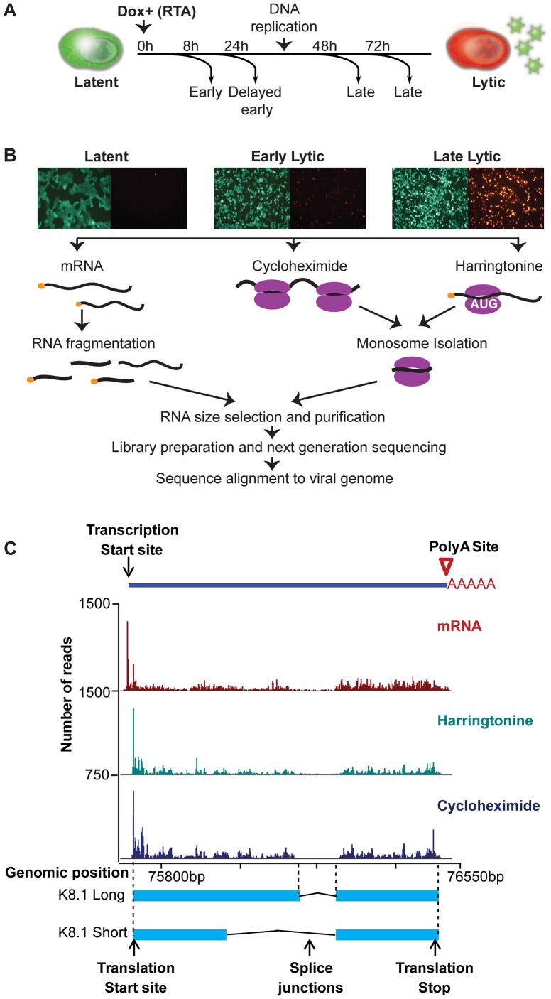
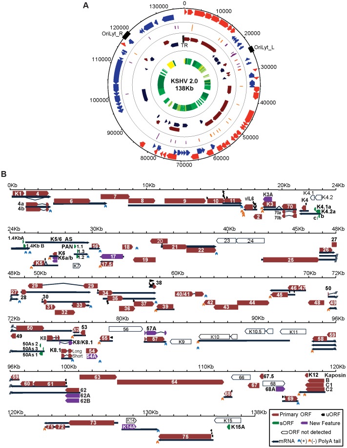
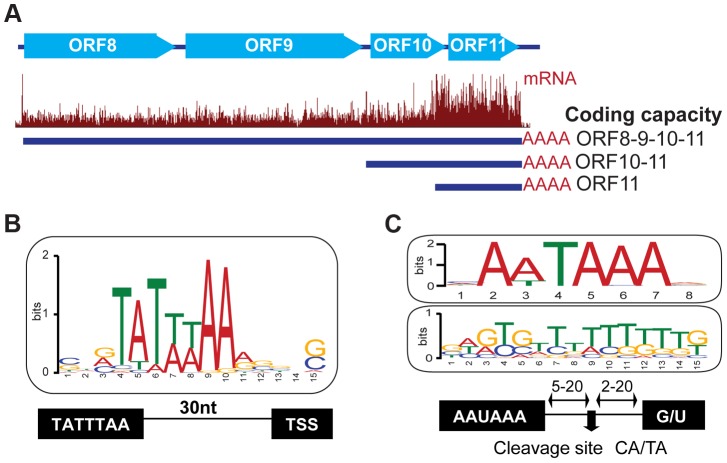
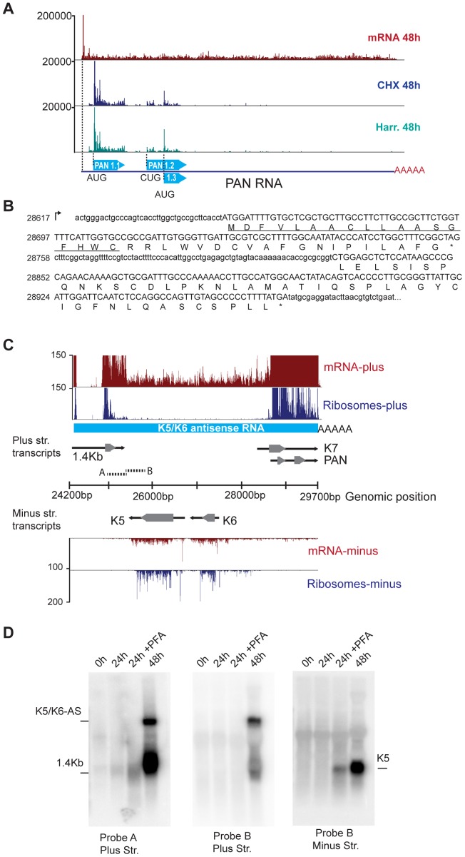
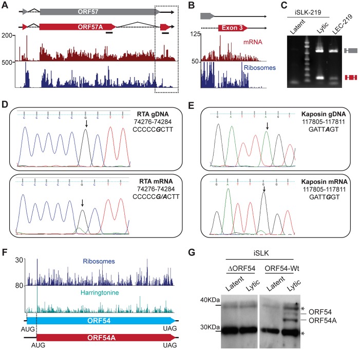
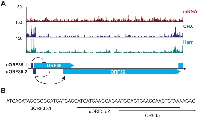
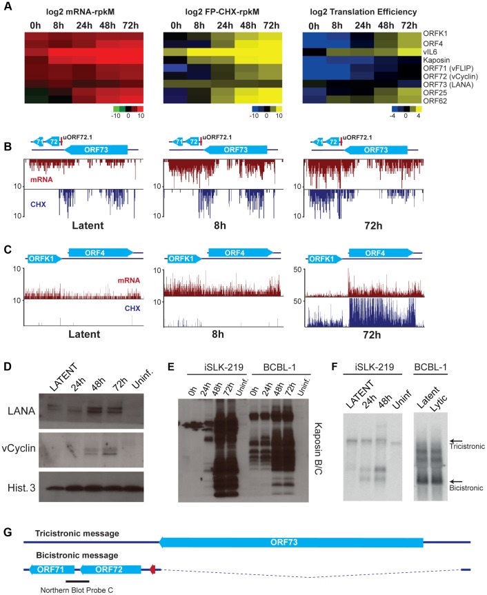
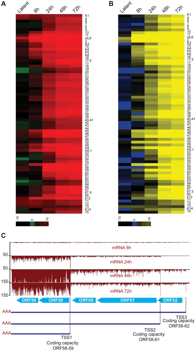
Productive herpesvirus infection requires a profound, time-controlled remodeling of the viral transcriptome and proteome. To gain insights into the genomic architecture and gene expression control in Kaposi's sarcoma-associated herpesvirus (KSHV), we performed a systematic genome-wide survey of viral transcriptional and translational activity throughout the lytic cycle. Using mRNA-sequencing and ribosome profiling, we found that transcripts encoding lytic genes are promptly bound by ribosomes upon lytic reactivation, suggesting their regulation is mainly transcriptional. Our approach also uncovered new genomic features such as ribosome occupancy of viral non-coding RNAs, numerous upstream and small open reading frames (ORFs), and unusual strategies to expand the virus coding repertoire that include alternative splicing, dynamic viral mRNA editing, and the use of alternative translation initiation codons. Furthermore, we provide a refined and expanded annotation of transcription start sites, polyadenylation sites, splice junctions, and initiation/termination codons of known and new viral features in the KSHV genomic space which we have termed KSHV 2.0. Our results represent a comprehensive genome-scale image of gene regulation during lytic KSHV infection that substantially expands our understanding of the genomic architecture and coding capacity of the virus.


Conflict of interest statement

The authors have declared that no competing interests exist.

Figures

Figure 1
Figure 1. High-resolution mapping of KSHV genomic features.
(A–B) Strategy for identification of transcripts and ribosome footprints in iSLK-219 cells. Latent iSLK-219 cells were induced to enter the lytic cycle by expression of the doxycycline-inducible transcription factor RTA for 8, 24, 48 and 72 hr. mRNA and ribosome footprints were isolated from cells and the purified RNA was deep sequenced. (C) Ribosome occupancy and mRNA profiles of the late lytic gene K8.1 at 48 hr post induction (hpi).
Figure 2
Figure 2. KSHV 2.0: A high-resolution functional genome map of KSHV.
(A) Circular map of the KSHV genome. The outer circle represents the genomic coordinates. The concentric circles illustrate, from the outermost to the innermost: viral ORFs (Red sense, blue antisense), sORFs (purple) and uORFs (orange), transcripts (maroon sense, dark blue antisense) and timing of expression (from yellow to dark green: latent, early lytic, delayed early lytic, late lytic). (B) Linear map of the KSHV genome showing the details of genomic features illustrated in A. ORF29, ORF32 and ORF36 are depicted as annotated ORFs as we detected the presence of ribosomes in their coding sequences. The exact boundaries of translation for these ORFs could not be determined. See also Tables 1, 2 and 3.
Figure 3
Figure 3. Host cis-regulatory elements are conserved in viral transcripts.
(A) mRNA-Seq of the locus encoding the ORF8/9/10 and 11 genes at 72 hr post reactivation. Three TSS sharing a common PAS were mapped within this polycistronic locus. (B) Sequence analysis of 50 nucleotides flanking the predicted TSS site for 49 viral genes. Note the presence of the TATA box 30 bp upstream of the TSS. (C) Sequence analysis of 30 nt flanking the 42 polyA sites mapped in KSHV. Note the conserved AA/UUAAA motif 5–20 nt upstream (top panel), and the GU rich region (middle panel) 5–20 nt downstream of the cleavage site. See also Tables S1 and S2.
Figure 4
Figure 4. mRNA-Seq reveals ribosome-protected non-coding RNAs in KSHV.
(A) The PAN transcript is protected by ribosomes during the lytic cycle. mRNA-Seq and Ribo-Seq of the PAN gene at 48 hr post reactivation. Ribo-Seq in cells treated with CHX (blue) or Harr (teal) show the accumulation of ribosomes on three distinct coding regions of this transcript. (B) PAN codes for three putative sORFs, PAN1.1 (37aa), PAN1.2 (44aa), and PAN1.3 (25aa), which can be identified as ribosome-populated regions (in capital letters). The predicted signal peptide in PAN1.1 is underlined. (C) The K5/K6 antisense transcript is devoid of nucleosomes following lytic reactivation. mRNA-Seq and Ribo-Seq of the genomic region from 24200 bp to 29700 bp at 48 hr post induction. The light blue solid line corresponds to the K5/K6 antisense RNA. Solid black arrows indicate transcripts and grey arrowheads indicate coding regions. (D) Northern blot for the same region depicted in (C). The A and B probes are indicated as short broken lines in (C). PFA: phosphonoformate. See also Figure S5 and Table S3.
Figure 5
Figure 5. KSHV employs multiple strategies to expand and regulate its coding repertoire.
(A–C) Novel splice variant of ORF57 detected by mRNA-Seq. Tracks for mRNA-Seq and Ribo-Seq in CHX treated cells (48 hpi) show the new, shorter-splice variant of ORF57, ORF57A (red box). Splicing of the second intron in ORF57 removes the UAA stop codon, resulting in the generation of a 33 aa C-terminal exon. (B) Zoom in of the region highlighted in (A). Note that ribosomes accumulate downstream of the ORF57 stop codon, indicating that the new exon may be translated. (C) Verification of ORF57A splicing by end-point PCR in lytic iSLK-219 (72 hpi) and LEC-219 cells. The primers used for amplification flank the intron boundaries and are shown in panel (A) as solid black lines. The high molecular weight product (∼800 bp) corresponds to the unspliced form of ORF57. The low molecular weight product (165 bp), corresponds to ORF57A, from which the second intron has been removed. (D–E) mRNA editing in the RTA and Kaposin transcripts. Sanger sequencing of RTA (D) and Kaposin (E) genomic DNA (top panel) and cDNAs (48 hpi) (bottom panel) from iSLK-219 cells. The edited nucleotide is bold, italic font. (F–G) ORF54 translation starts from two alternative initiation codons. (F) Ribo-Seq tracks for cycloheximide (CHX blue) and harringtonine (Harr teal) treated cells at 72 hpi. The dotted lines indicate the two translation initiation sites used for ORF54 translation. (G) Immunoblot for ORF54 in latent and lytic (48 hpi) iSLK cells, infected with Wt KSHV or an ORF54 knock-out virus (ΔORF54). *Non-specific product. See also Figures S6, S7 and S8.
Figure 6
Figure 6. KSHV encodes functional uORFs.
(A) mRNA-Seq and Ribo-Seq tracks (CHX and Harr) show the accumulation of ribosomes in the regions of uORF35.1 and uORF35.2. These uORFs regulate the expression of ORF35 and ORF36 (adapted from [57]). (B) Start and coding sequences for uORF35.1, uORF35.2 and ORF35. See also Table S7.
Figure 7
Figure 7. Gene expression is limited during latency in iSLK-219 cells.
(A) Log2 of normalized mRNA-Seq, Ribo-Seq counts (reads per kilobase million, rpkM) and translation efficiency (TE = Ribo-seq rpkM/mRNA-seq rpkM) for the latent transcripts and two late lytic ORFs (ORF25 and ORF62). Note the low TE for the latent genes at 0 h and 8 h. (B–C) Ribosome occupancy of latent transcripts changes with time. mRNA-seq (red) and Ribo-seq (CHX blue) for (B) the latency locus and (C) the K1-ORF4 region. (D–E) Immunoblot for the products of the latent transcripts (D) LANA and vCyclin, and (E) Kaposin. (F) Northern blot for the tricistronic (LANA-vCyclin-vFLIP) and the bicistronic (vCyclin-vFLIP) illustrated in (G). mRNA (100 ng-iSLK-219) or total RNA (10 ug-BCBL-1) were probed for vCyclin and vFLIP (see black bar in G). (G) Schematic of the tricistronic and bicistronic transcripts in the major latency locus.
Figure 8
Figure 8. Lytic reactivation is subject to a transcriptional timer.
(A–B) Log2 of normalized mRNA-Seq (A) and Ribo-Seq (B) counts (reads per kilobase million, rpkM) was calculated for the primary ORFs. The log2 scores reflect mRNA-expression (mRNA-seq) or ribosome occupancy (Ribo-seq) for each gene. The ORFs are organized by genomic position from ORFK1 to ORF75. The ORFs that were not annotated in our study have not been included in the plots (C) mRNA-Seq for the ORF58–62 locus. Three TSS sharing a single PAS were mapped in this region. Note the timing of expression for each of the transcripts.

References

    1. Cesarman E, Knowles DM (1999) The role of Kaposi's sarcoma-associated herpesvirus (KSHV/HHV-8) in lymphoproliferative diseases. Seminars in cancer biology 9: 165–174. - PubMed
    1. Ganem D (2010) KSHV and the pathogenesis of Kaposi sarcoma: listening to human biology and medicine. The Journal of clinical investigation 120: 939–949. - PMC - PubMed
    1. Chang Y, Cesarman E, Pessin MS, Lee F, Culpepper J, et al. (1994) Identification of herpesvirus-like DNA sequences in AIDS-associated Kaposi's sarcoma. Science (New York, NY) 1865–1869. - PubMed
    1. Russo JJ (1996) Nucleotide sequence of the Kaposi sarcoma-associated herpesvirus (HHV8). Proceedings of the National Academy of Sciences 93: 14862–14867. - PMC - PubMed
    1. Gottwein E (2012) Kaposi's Sarcoma-Associated Herpesvirus microRNAs. Frontiers in microbiology 3: 165. - PMC - PubMed

Publication types

LinkOut - more resources