. 2014 Jan;10(1):e1003876.
doi: 10.1371/journal.ppat.1003876. Epub 2014 Jan 16.

Phosphoenolpyruvate carboxylase identified as a key enzyme in erythrocytic Plasmodium falciparum carbon metabolism

Affiliations

Phosphoenolpyruvate carboxylase identified as a key enzyme in erythrocytic Plasmodium falciparum carbon metabolism

Janet Storm et al. PLoS Pathog. 2014 Jan.

Abstract

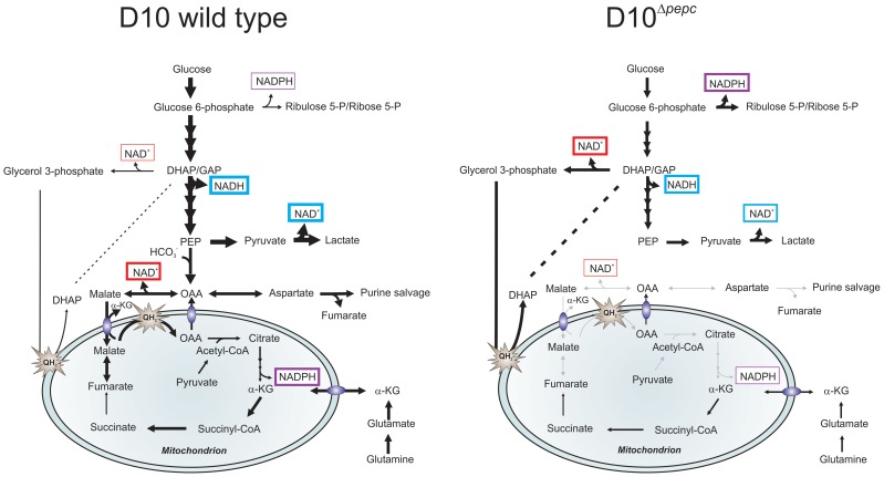
Phospoenolpyruvate carboxylase (PEPC) is absent from humans but encoded in the Plasmodium falciparum genome, suggesting that PEPC has a parasite-specific function. To investigate its importance in P. falciparum, we generated a pepc null mutant (D10(Δpepc) ), which was only achievable when malate, a reduction product of oxaloacetate, was added to the growth medium. D10(Δpepc) had a severe growth defect in vitro, which was partially reversed by addition of malate or fumarate, suggesting that pepc may be essential in vivo. Targeted metabolomics using (13)C-U-D-glucose and (13)C-bicarbonate showed that the conversion of glycolytically-derived PEP into malate, fumarate, aspartate and citrate was abolished in D10(Δpepc) and that pentose phosphate pathway metabolites and glycerol 3-phosphate were present at increased levels. In contrast, metabolism of the carbon skeleton of (13)C,(15)N-U-glutamine was similar in both parasite lines, although the flux was lower in D10(Δpepc); it also confirmed the operation of a complete forward TCA cycle in the wild type parasite. Overall, these data confirm the CO2 fixing activity of PEPC and suggest that it provides metabolites essential for TCA cycle anaplerosis and the maintenance of cytosolic and mitochondrial redox balance. Moreover, these findings imply that PEPC may be an exploitable target for future drug discovery.


Conflict of interest statement

The authors have declared that no competing interests exist.

Figures

Figure 1
Figure 1. Gene replacement of pepc.
(A) Schematic representation of the endogenous pepc locus, the pCC4-Δpepc plasmid and the pepc locus following integration by double crossover recombination of pCC4-Δpepc (D10Δpepc). SpeI restriction sites for diagnostic Southern blot analyses and sizes of the expected DNA fragments are indicated. Two regions homologous to the 5′ (Δpepc 5′) and 3′ (Δpepc 3′) end of pepc present in pCC4-Δpepc recombine with the endogenous pepc locus and part of the pepc gene is replaced with the positive selectable marker blasticidin-S-deaminase (bsd), which is under control of the calmodulin promoter (cam 5′) and the histine rich protein 2 3′ UTR (hrp2 3′). The plasmid contains the negative selectable marker cytosine deaminase (CD), which is under control of the heat shock protein 86 promotor (hsp86 5′) and the P. berghei dihydrofolate reductase 3′ UTR (PbDT 3′), and is lost upon double crossover recombination. (B) Southern blot of SpeI-digested genomic DNA of wild type parasites and parasites transfected with pCC4-Δpepc, cultured in routine medium or malate medium, probed with the 5′ end of pepc. Cycle 1 (c1) refers to the first parasites resistant to blasticidin and 5-fluorocytosine (5-FC), while cycle 2 (c2) are 5-FC-resistant parasites after 1 additional drug selection cycle. Integration of the plasmid only occurred when the transfectants were cultured in malate medium (diagnostic fragment: 5.0 kb). The derived clones D10Δpepc-1 and D10Δpepc-2 are shown in lane 5 and 6. In routine medium no integration was observed, only fragments corresponding to endogenous pepc (7.1 kb), plasmid (6.2 kb and 2.1 kb) and five fragments of unknown identity (*) were detected.
Figure 2
Figure 2. Growth phenotype of D10Δ pepc and rescue by malate and fumarate.
(A) Growth of D10 in routine medium lacking malate and D10Δpepc in malate or routine medium was followed for 14 days. The cultures were diluted 1∶5 on even days. The means ± S.D. of two (D10) and three (D10Δpepc) replicates are shown. (B and C) D10 and D10Δpepc were cultured in routine medium for 9 days, synchronised, diluted to 1% parasitaemia and cultured for 5 days in medium supplemented with increasing concentrations of malate (0.5–5 mM) (B) or a range of metabolites (C). The cultures were diluted 1∶5 on day 3. The parasitaemia on day 5 was determined and multiplied with the dilution factor to give the accumulated parasitaemia. Data are means ± S.E.M. of at least 3 independent experiments, each done in triplicate (for succinate and citrate, means ± S.D. of 2 experiments done in triplicate). The asterisks indicate a statistically significant increase in parasitaemia compared to routine medium (p<0.05). Other apparent changes were not statistically significant. The final concentrations of metabolites used in (C) were: 0.5 mM citrate, 5 mM all others.
Figure 3
Figure 3. I-TASSER homology model of P. falciparum PEPC.
Structural alignment of an I-TASSER predicted model for PfPEPC with the top structural analogue EcPEPC is shown. PfPEPC is represented in a ribbon diagram where α-helices are in green, β-sheets are in yellow and coils are in grey. The EcPEPC ribbon diagram is represented completely in blue. Coincident structures are dual-coloured. Based on the homology modelling, three regions of PfPEPC were found not to overlap with EcPEPC. Two of these regions are visible in (A), (Glu363 to Ser391 and Gln954 to Gly1148). The structure rotated to the left is displayed in (B) to show the third non-homologous region (Met1 to Cys46). The C-terminus of PfPEPC appears to be entirely non-homologous with EcPEPC presumably because of a long Plasmodium-specific insertion (from Gln954) that separates the terminal 9 amino acid sequence, which is highly conserved in the parasite protein, from the core structure (Fig. S2).
Figure 4
Figure 4. Efficacy of L-cycloserine, DSM190 and atovaquone against D10 and the D10Δ pepc in routine and malate media.
The effect of L-cycloserine (A), DSM190 (B) and atovaquone (C) on parasite viability. D10Δpepc was maintained for 9 days in routine medium prior to the experiment.
Figure 5
Figure 5. Metabolite labelling from 13C-U-D-glucose.
(A) Schematic representing glucose utilisation in P. falciparum based on the utilisation of 13C-U-D-glucose and distribution of 13C carbons into metabolic intermediates. (B) The parasites perform glycolysis such that triose 3-phosphates, phosphoenolpyruvate (PEP) and lactate had very extensive labelling with 13C from 13C-U-D-glucose (all are three carbon compounds, hence exist mainly as 13C-3 molecules as rapidly generated from glucose by glycolysis). 13C is also incorporated into malate, fumarate and aspartate through the action of PEPC (and hence the 13C-3 molecules) as well as the TCA metabolite citrate by the action of a PDH-like enzyme , in D10 parasites. Citrate is formed from OAA (occurring as 13C-3 molecule as well as unlabelled) and acetyl-CoA (presumably mainly 13C-2 molecules as generated from pyruvate) and so was present as 13C-2 and 13C-5 molecules as well as unlabelled. 13C-labelled malate, fumarate, asparate and citrate were absent from samples of D10Δpepcpepc). 13C-U-D-glucose was also fed into the PPP intermediates ribulose 5-P/ribose 5-P and sedoheptulose 7-P (compounds with 5 and 7 carbons, respectively, hence the extensive presence as 13C-5 and 13C-7 molecules) and used to generate glycerol 3-phosphate (also a 3-carbon molecule hence the 13C-3 labelling). Legend: UL, unlabelled; 13C-1: one carbon atom labelled; 13C-2 to 13C-7: two to seven carbon atoms labelled; D1090, D10-infected RBC concentrated to 90% parasitaemia; D106, D10-infected RBC at 6% parasitaemia. (C) Relative levels of intracellular metabolites in D10Δpepcpepc) and D10. Abbreviations: α-KG, α-ketoglutarate; DHAP, dihydroxyacetone phosphate; GAP, glyceraldehyde 3-phosphate; OAA, oxaloacetate; PEP, phosphoenolpyruvate; PPM, parasite plasma membrane; PVM, parasitophorous vacuole membrane; QH2, ubiquinol.
Figure 6
Figure 6. Metabolite labelling from 13C-bicarbonate.
(A) Schematic representing bicarbonate utilisation in P. falciparum by determining the incorporation of the single 13C isotope into metabolic intermediates. (B) 13C-bicarbonate is incorporated into malate and fumarate and also to a lesser extent into aspartate and citrate. The natural relative abundance of 13C-1 label in metabolites was not subtracted from the data shown. This accounts for ∼1.1% of 13C-1 for each carbon of the metabolites in all samples analysed including those of D10Δpepcpepc) and RBC, which show no specific additional incorporation of 13C-bicarbonate into their carbon skeleton. Thus the specific incorporation into the metabolites is most usefully assessed by comparing the levels in parasite-infected cells with that measured in the RBC samples (which reflects the natural abundance of the isotope). Abbreviations: as in Figure 5 and m, spent medium samples.
Figure 7
Figure 7. Hypothetical model of the contributions PEPC and adaptations of D10Δ pepc to compensate for lack of PEPC.
The width of arrows is indicative of the fluxes through the pathways. Glucose used by wild type parasites is phosphorylated into glucose 6-phosphate, which is either catabolised in the glycolytic pathway to generate lactate or is used in the PPP to provide NADPH and 5 and 7-carbon sugars for downstream metabolic processes. During glycolysis, NAD+ reduction and NADH oxidation (blue boxes) are balanced in the complete glycolytic sequence itself, but some metabolites are used in other ways. Glycerol 3-phosphate dehydrogenase and especially MDH, converting OAA (the product of CO2 fixation by PEPC) to malate, play roles in maintenance of redox balance in the cytoplasm (red boxes). Pyruvate is metabolised not only into lactate but is also transferred into the mitochondrion where it is oxidised into acetyl-CoA by a PDH-like dehydrogenase , . Malate is transferred into the mitochondrion via a malate∶α-ketoglutarate (αKG) antiporter and enters TCA metabolism leading to OAA formation in the organelle, this oxidation transfers reducing equivalents into the mtETC (QH2). OAA may leave the mitochondrion, thus completing a parasite-specific form of a malate shuttle, or be converted to citrate as part of canonical TCA metabolism. Deletion of pepc leads to an imbalance in redox metabolism in both cytosol and presumably also mitochondrion. D10Δpepc apparently adapted to this with increased generation of glycerol 3-phosphate with concomitant NADH oxidation, thus compensating partially for the loss of downstream generation of NAD+ through MDH. Increased flux through oxidative PPP is a mechanism for generating NADPH which may be compensating for the reduced flux through TCA metabolism and therefore a reduced generation of NADPH at the IDH step in D10Δpepc(purple boxes). The reduction in flux to lactate in D10Δpepc reflects the severely reduced growth phenotype of D10Δpepc. The flux from glutamine via glutamate to α-ketoglutarate and, within the mitochondrion, to succinate and, at an apparently lower flux, to fumarate and malate is shown, with α-ketoglutarate as an additional entry point into TCA metabolism. The flux through these metabolites is lower in D10Δpepc compared with D10, reflecting the general growth defect of D10Δpepc and also its reduced consumption of glutamine, although the pathway in which the carbon skeleton is incorporated is not dramatically affected.

Similar articles

Cited by

References

    1. WHO, (2012) World Malaria Report 2012, World Health Organisation, Geneva, Switzerland, 59.
    1. Hay SI, Guerra CA, Gething PW, Patil AP, Tatem AJ, et al. (2009) A world malaria map: Plasmodium falciparum endemicity in 2007. PLoS Med 6: e1000048. - PMC - PubMed
    1. MacRae JI, Dixon MW, Dearnley MK, Chua HH, Chambers JM, et al. (2013) Mitochondrial metabolism of sexual and asexual blood stages of the malaria parasite Plasmodium falciparum . BMC Biol 11: 67. - PMC - PubMed
    1. Foth BJ, Stimmler LM, Handman E, Crabb BS, Hodder AN, et al. (2005) The malaria parasite Plasmodium falciparum has only one pyruvate dehydrogenase complex, which is located in the apicoplast. Mol Microbiol 55: 39–53. - PubMed
    1. Pei Y, Tarun AS, Vaughan AM, Herman RW, Soliman JM, et al. (2010) Plasmodium pyruvate dehydrogenase activity is only essential for the parasite's progression from liver infection to blood infection. Mol Microbiol 75: 957–971. - PubMed

Publication types

MeSH terms