Clinical Trial
. 2014 Jan;10(1):e1003885.
doi: 10.1371/journal.ppat.1003885. Epub 2014 Jan 16.

Malaria-induced NLRP12/NLRP3-dependent caspase-1 activation mediates inflammation and hypersensitivity to bacterial superinfection

Affiliations
Clinical Trial

Malaria-induced NLRP12/NLRP3-dependent caspase-1 activation mediates inflammation and hypersensitivity to bacterial superinfection

Marco A Ataide et al. PLoS Pathog. 2014 Jan.

Erratum in

  • PLoS Pathog. 2014 Jun;10(6):e1004258

Abstract

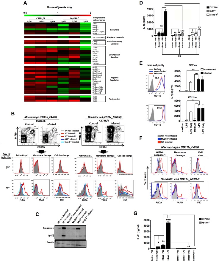
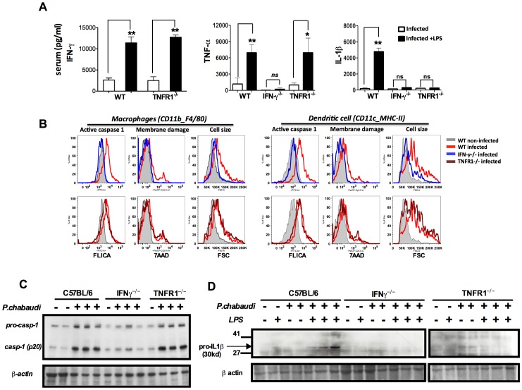
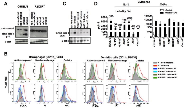
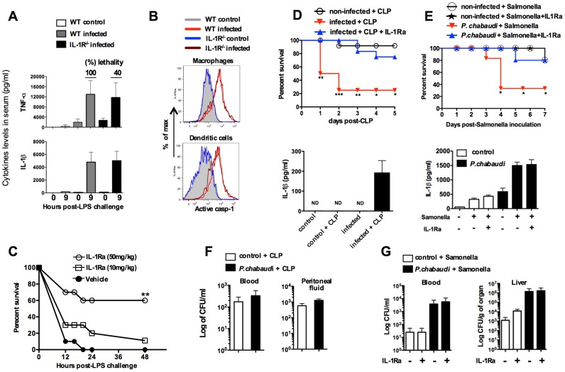
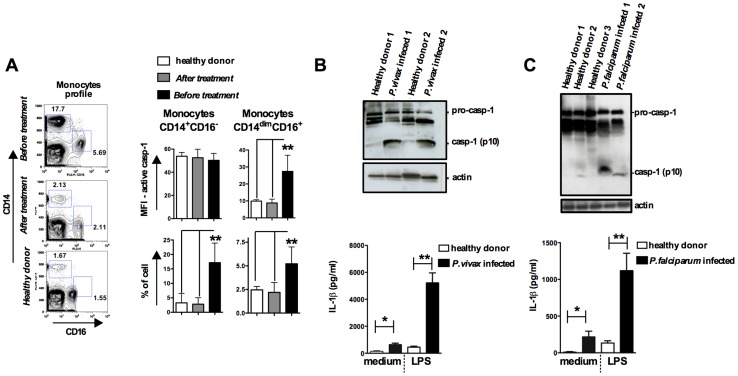
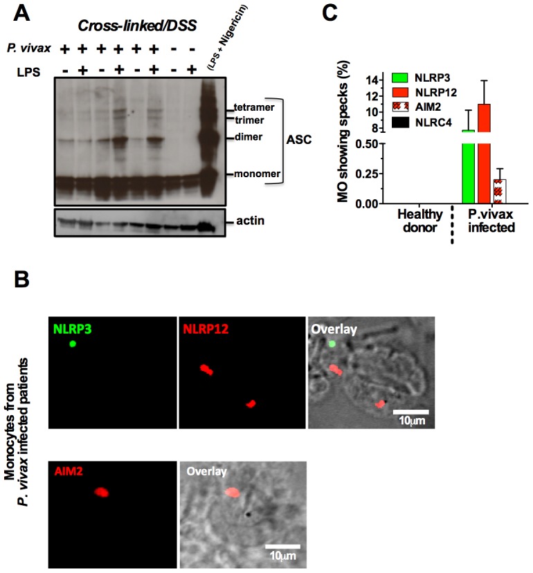
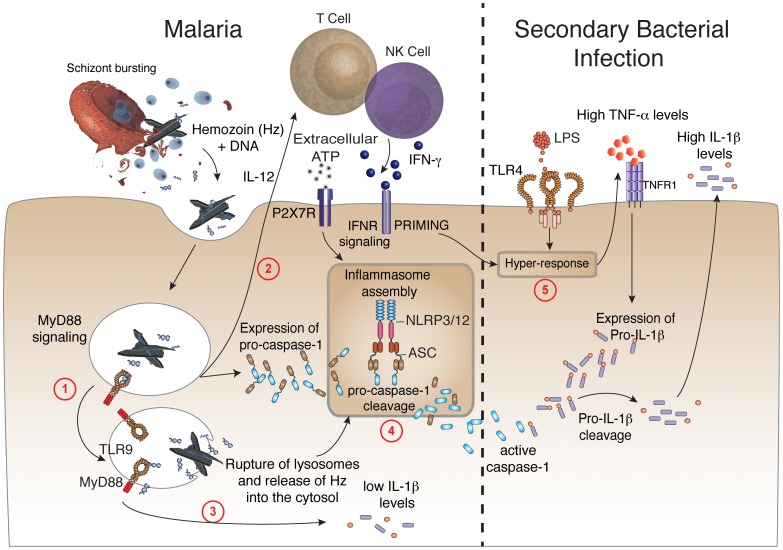
Cyclic paroxysm and high fever are hallmarks of malaria and are associated with high levels of pyrogenic cytokines, including IL-1β. In this report, we describe a signature for the expression of inflammasome-related genes and caspase-1 activation in malaria. Indeed, when we infected mice, Plasmodium infection was sufficient to promote MyD88-mediated caspase-1 activation, dependent on IFN-γ-priming and the expression of inflammasome components ASC, P2X7R, NLRP3 and/or NLRP12. Pro-IL-1β expression required a second stimulation with LPS and was also dependent on IFN-γ-priming and functional TNFR1. As a consequence of Plasmodium-induced caspase-1 activation, mice produced extremely high levels of IL-1β upon a second microbial stimulus, and became hypersensitive to septic shock. Therapeutic intervention with IL-1 receptor antagonist prevented bacterial-induced lethality in rodents. Similar to mice, we observed a significantly increased frequency of circulating CD14(+)CD16(-)Caspase-1(+) and CD14(dim)CD16(+)Caspase-1(+) monocytes in peripheral blood mononuclear cells from febrile malaria patients. These cells readily produced large amounts of IL-1β after stimulation with LPS. Furthermore, we observed the presence of inflammasome complexes in monocytes from malaria patients containing either NLRP3 or NLRP12 pyroptosomes. We conclude that NLRP12/NLRP3-dependent activation of caspase-1 is likely to be a key event in mediating systemic production of IL-1β and hypersensitivity to secondary bacterial infection during malaria.


Conflict of interest statement

The authors have declared that no competing interests exist.

Figures

Figure 1
Figure 1. Caspase-1 activation, IL-1β production and pyroptosis in splenic macrophages and DCs from P. chabaudi infected mice.
(A) Gene expression was determined in splenocytes of 3 C57BL/6 or MyD88−/− mice at 6 days post-infection over 3 non-infected controls by Microarray analysis. (B and F) Splenocytes from C57BL/6, ASC−/−, Casp-1−/− or MyD88−/− mice were stained and analyzed by FACs to gate macrophages (CD11b+F4/80+) and DCs (CD11c+MHC-II+). Active caspase-1 was evaluated by FLICA reagent, membrane integrity by nuclei staining with 7AAD, and cell size change by shift on FSC axis. The results are representative from 3 experiments that yield similar results. (C) On day 7 post-infection splenocytes from C57BL/6, ASC−/−, Casp-1−/− and MyD88−/− mice were lysed by RIPA buffer and analyzed by Western blot employing an anti-caspase-1 antibody. A faint band of similar molecular weight of active caspase-1 corresponds to IgG light chain is seen in the infected ASC−/− and Casp-1−/− mice. (D and G) At 7 days post-infection mice were inoculated intravenously with 10 µg of LPS per mouse, and 9 hours later, sera was collected for measuring the levels of circulating IL-1β. The average levels of IL-1β in control and infected mice, before LPS challenge, were 64.2 and 434.2 pg/ml in figure D, and 82.2 and 602.4 pg/ml in figure G. These results are the means + SEM of 10–15 animals from 3 independent experiments that yield similar results. (E) CD11c+ and CD11b+ cells highly purified from spleens of C57BL/6 mice at day post-infection were cultured with LPS (1 µg/ml) and supernatants harvested 18 h later to measure the levels of IL-1β. As positive control we used the purified cells stimulated with LPS at same concentration followed by nigericin at 5 µM. Significant differences are indicated by *p<0.01, **p<0.001 and ***p<0.0005 obtained in the Mann-Whitney test.
Figure 2
Figure 2. Both endogenous IFN-γ and TNF-α are required for IL-1β production in mice infected with P. chabaudi.
(A) Mice were challenged with 10 µg of LPS, at 7 days post-infection, and sera collected 9 hours later for cytokine measurements. These results are means + SEM of 10 animals from 2 independent experiments. Significant differences are *p = 0.034 and **p = 0.001 as indicated by the Mann-Whitney test. (B) Active caspase-1, membrane integrity and cell size were assessed in macrophages (CD11b+F4/80+) and DCs (CD11c+MHC-II+) by flow cytometry, employing the FLICA reagent, nuclei staining with 7AAD, and the shift on FSC axis, respectively. These results presented in figures are representative of 2 experiments. (C) Splenocytes lysates were obtained from mice at 7 days post-infection and used in Western blot analysis. A faint band of similar molecular weight of active caspase-1 that corresponds to IgG light chain is seen in the uninfected controls or infected IFN-γ−/− mice. These results presented in figures are representative of 2 experiments. (D) Two hours after LPS-challenge, splenocytes lysates were harvested to evaluate expression of pro-IL-1β. These results are representative of 3 independent experiment that yielded similar results.
Figure 3
Figure 3. NLRP3/NLRP12-dependent activation of caspase-1 and pyroptosis in mice infected with P. chabaudi.
(A) At 9 days post-infection, splenocytes from C57BL/6, and P2X7R−/− mice were lysed and analyzed by Western blot employing caspase-1-specific antibody. (B) At 7 days post-infection, active caspase-1 by FLICA reagent, membrane integrity by propidium iodide, and cell size change by shift on FSC axis were assessed in splenic macrophages (CD11b+F4/80+) and DCs (CD11c+MHC-II+). (C) Splenocytes lysates from mice at 7 days post-infection were used to detected active caspase-1 by Western blot. A faint band of similar molecular weight of active caspase-1 that corresponds to IgG light chain is seen in the uninfected controls or in various infected knockout mice. The results presented in figures 3A, 3B and 3C are representative of 2 experiments that yield similar results. (D) A LPS dose of 10 µg/mouse was given intravenously at 7 days post-infection with P.chabaudi and sera collected 9 hours later, for measuring IL-1β and TNF-α levels. The numbers within parenthesis indicate the percentage of lethality 48 hours after LPS challenge (10 µg/mouse). The levels of IL-1β measured in the sera of infected C57BL/6, NLRP3−/−, NLRP12, ASC−/− and Casp-1−/− were not different. The results are means + SEM of 10 mice from 2 independent experiments. Significant differences are indicated by **p<0.001 obtained in the Mann-Whitney test.
Figure 4
Figure 4. Treatment with IL-1RA prevents lethality in mice infected with P. chabaudi and challenged with a secondary bacterial infection.
(A) At 7 days post-infection, mice were challenged with 10 µg of LPS and serum samples collected 9 hours later for cytokine measurements. The numbers within parenthesis indicate the percentage of lethality 24 hours after low dose (10 µg/mouse) LPS challenge. (B) Splenic macrophages (CD11b+F4/80+) and DCs (CD11c+MHC-II+) from mice at 7 days post-infection were stained with FLICA reagent in order to detect active caspase-1. (C) At day 7 post-infection the mice were treated with IL-1RA (anakinra) immediately prior to LPS challenge. Lethality was assessed from 12 to 48 hours post-LPS challenge. (D) At 7 days post-infection with P. chabaudi, sub-lethal sepsis was induced by CLP. A group of mice received treatment with IL-1RA (100 mg/kg/day) beginning 24 hours before the CLP procedure. Levels of circulating IL-1β were measured 24 hs after CLP. (E) Mice received peroral challenge with 108 of Salmonella typhimurium at 5 days post-infection with P. chabaudi. A group of mice received treatment with IL-1RA (100 mg/kg/day) beginning 48 hours after bacterial challenge. The levels of circulating IL-1β were measured at 3 days post-Salmonella challenge. (F) Translocation of aerobic bacteria was quantified 24 hours after the CLP procedure. (G) Translocation of S. typhimurium was quantified 3 days after peroral challenge. We used 5 to 8 mice per group and results shown are representative of 2 independent experiments. Significant differences are *p<0.01, **p<0.005 ***p<0.001 obtained in a Chi-square test.
Figure 5
Figure 5. Monocytes are the major source of active caspase-1 during malaria.
PBMCs were obtained from either P. vivax or P. falciparum malaria patients as well as from healthy donors. (A) The gate was set based on monocytes profile in PBMCs from P. vivax infected patients. PBMCs were stained with anti-CD14 and anti-CD16 antibodies to determine the presence of different monocyte subsets. These cells were gated based on FSC and SSC to avoid neutrophil contamination. When the CD14dimCD16+ gate was moved down in PBMCs from healthy donors or from malaria patients after treatment, we did not detect any active caspase-1. The bar graphs show the flow cytometry analysis of PBMCs from five P. vivax infected subjects before and after malaria treatment primaquine and chloroquine, as well as eight healthy donors. To determine the median fluorescence intensity (MFI) and frequency of (CD14+CD16) and (CD14dimCD16+) that are active caspase-1, we used the FLICA assay. (B – top panel) Active caspase-1 (p10) was detected in lysates of PBMCs from P. vivax or (C – top panel) P. falciparum infected individuals by Western blot. (B and C – bottom panel) PBMCs were stimulated with LPS (100 ng/ml) for 24 hours, and levels of IL-1β assessed in the culture supernatants by ELISA. Significant differences are *p<0.05 and **p<0.005 as indicated by the unpaired t test with Welch correction or Mann-Whitney test when a normality test failed.
Figure 6
Figure 6. NLRP3/NLRP12 containing inflammasomes and caspase-1 activation in PBMCs from P. vivax malaria patients.
(A) PBMCs derived from P. vivax malaria patients and healthy donors were lysed, cross-linked by treatment with disuccinimidyl suberate , and ASC oligomerization assessed by Western blot analysis. PBMCs from a healthy donor stimulated with LPS and nigericin were used as positive control. . (B) NLRP3, NLRP12 and AIM2 containing inflammasomes (specks) in monocytes from P. vivax malaria patients were visualized in a confocal microscope. (C) The bar graphs show the frequency of specks in monocytes derived from P. vivax malaria patients. We saw no specks on cells from healthy donors or cells from malaria patients incubated with the secondary antibody only. See also Figure S6.
Figure 7
Figure 7. Malaria-induced NLRP12/NLRP3-dependent caspase-1 activation mediates IL-1β and hypersensitivity to bacterial superinfection.
Step 1 –Phagocytes internalize Plasmodium DNA bound to hemozoin that activates TLR9 and the adaptor molecule named MyD88. Step 2 – MyD88 signaling triggers the expression of IL-12, which will initiate the production of IFN-γ by T lymphocytes and NK cells. Step 3 – Low levels of caspase-1-independent IL-1β induced by malaria infection. Step 4 – IFN-γ priming and MyD88 signaling in phagocytes will lead to enhanced expression of pro-caspase-1. K+ efflux as well as rupture (by hemozoin crystals) and release of lysosome contents will induce the assembly of ASC, NLRP3 and NLR12 inflammasomes and promote cleavage of pro-caspase-1. Step 5 - Bacterial superinfection triggers expression of high pro-IL-1β levels, in a TNF-α-dependent manner. Pro-IL-1β will be cleaved by active caspase-1 generated on step 4. Upon secondary bacterial infection, the malaria-primed macrophages will release deleterious amounts of IL-1β.

References

    1. Murray CJ, Rosenfeld LC, Lim SS, Andrews KG, Foreman KJ, et al. (2012) Global malaria mortality between 1980 and 2010: a systematic analysis. Lancet 379: 413–431. - PubMed
    1. Mueller I, Galinski MR, Baird JK, Carlton JM, Kochar DK, et al. (2009) Key gaps in the knowledge of Plasmodium vivax, a neglected human malaria parasite. Lancet Infect Dis 9: 555–566. - PubMed
    1. Miller LH, Ackerman HC, Su XZ, Wellems TE (2013) Malaria biology and disease pathogenesis: insights for new treatments. Nat Med 19: 156–167. - PMC - PubMed
    1. Parroche P, Lauw FN, Goutagny N, Latz E, Monks BG, et al. (2007) Malaria hemozoin is immunologically inert but radically enhances innate responses by presenting malaria DNA to Toll-like receptor 9. Proc Natl Acad Sci U S A 104: 1919–1924. - PMC - PubMed
    1. Sharma S, DeOliveira RB, Kalantari P, Parroche P, Goutagny N, et al. (2011) Innate immune recognition of an AT-rich stem-loop DNA motif in the Plasmodium falciparum genome. Immunity 35: 194–207. - PMC - PubMed

Publication types

MeSH terms