. 2014 Jan;10(1):e1003893.
doi: 10.1371/journal.ppat.1003893. Epub 2014 Jan 16.

Glutamate utilization couples oxidative stress defense and the tricarboxylic acid cycle in Francisella phagosomal escape

Affiliations

Glutamate utilization couples oxidative stress defense and the tricarboxylic acid cycle in Francisella phagosomal escape

Elodie Ramond et al. PLoS Pathog. 2014 Jan.

Abstract

Intracellular bacterial pathogens have developed a variety of strategies to avoid degradation by the host innate immune defense mechanisms triggered upon phagocytocis. Upon infection of mammalian host cells, the intracellular pathogen Francisella replicates exclusively in the cytosolic compartment. Hence, its ability to escape rapidly from the phagosomal compartment is critical for its pathogenicity. Here, we show for the first time that a glutamate transporter of Francisella (here designated GadC) is critical for oxidative stress defense in the phagosome, thus impairing intra-macrophage multiplication and virulence in the mouse model. The gadC mutant failed to efficiently neutralize the production of reactive oxygen species. Remarkably, virulence of the gadC mutant was partially restored in mice defective in NADPH oxidase activity. The data presented highlight links between glutamate uptake, oxidative stress defense, the tricarboxylic acid cycle and phagosomal escape. This is the first report establishing the role of an amino acid transporter in the early stage of the Francisella intracellular lifecycle.


Conflict of interest statement

The authors have declared that no competing interests exist.

Figures

Figure 1
Figure 1. gadC inactivation affects intracellular survival and virulence.
Intracellular replication of wild-type F. novicida (WT) carrying the empty plasmid pKK214 (WT/pKK(−)), of the ΔgadC mutant (ΔgadC) and complemented strain (ΔgadC/pKK-gadC), and of the ΔFPI mutant (ΔFPI), was monitored in J774.1 macrophage-like cells (A); in THP-1 human macrophages (B); and in bone marrow-derived macrophages (C), over a 24 h-period. Results are shown as the average of log10 cfu mL−1 ± standard deviation. Each experiment was performed in triplicate. **, p<0.01 (as determined by the Student's t-test). Competition assays (D). A group of five female BALB/c mice were infected i.p. with a 1∶1 mixture of wild-type F. novicida and ΔgadC mutant strains (100 colony forming units (cfu) of each). The data represent the competitive index (CI) value for cfu of mutant/wild-type in the liver (L: black diamonds, left column) and spleen (S: black circles, right column) of each mouse, 48 h after infection. Bars represent the geometric mean CI value.
Figure 2
Figure 2. Stress sensitivity.
Exponential phase bacteria, diluted in TSB medium, were subjected: (A) to acidic stress (pH 5.5), or (B) to oxidative stress (500 µM H202). Exponential phase bacteria, diluted in chemically defined medium (CDM) (C), or CDM supplemented with 1 mM glutamate (D), were subjected to oxidative stress (500 µM H202). The bacteria were plated on chocolate agar plates at different times and viable bacteria were monitored 2 days after. Data are the average cfu mL−1 for three points. Experiments were realized twice. **, p<0.01 (as determined by the Student's t-test).
Figure 3
Figure 3. Subcellular localization of the ΔgadC mutant.
(A) Co-localization of wild-type F. novicida (1, 2, 3), ΔgadC (4, 5, 6) or ΔFPI mutant strain (7, 8, 9) with LAMP-1 was monitored by confocal microscopy, in J774.1 macrophage cells. Co-localization was monitored at 1 h (1, 4, 7), 4 h (2, 5, 8) and 10 h (3, 6, 9). Anti-Francisella antibody was used at a final dilution of 1∶500 and is shown in green. Anti-LAMP-1 antibody was used at a final dilution of 1∶100 and is shown in red. White arrowheads point to individual bacteria. Cell nuclei were labeled with DAPI (in blue). The images are representative from triplicate coverslips in three independent experiments. Scale bars at the bottom right of each panel correspond to 10 µM. (B) Quantification of co-localization between bacteria and LAMP-1 was obtained with Image J software. The graph results from the analysis of 4 different fields for each time of infection, in three independent experiments. **, p<0.01 (as determined by the Student's t-test). White bars, F. novicida U112 (WT); light grey bars, ΔgadC; dark grey bars, ΔFPI. (C) Transmission electron micrographs of thin sections of J774.1 macrophages, infected by wild-type F. novicida and ΔgadC mutant strains. Infections were monitored over a 10 h-period. At 10 h, active cytosolic multiplication of wild-type F. novicida was observed in most of the infected cells (1) whereas the ΔgadC mutant remains trapped into spacious phagosomes (2, 3, 4). Black arrowheads point to intact phagosomal membrane. (D) To evaluate the viability of intracellular Francisella, labeling with the cell-impermeant nucleic acid dye propidium iodide (PI) was performed. Confocal images of J774.1 cells, infected with wild-type F. novicida (1, 3) or ΔgadC mutant (2, 4) strain; after 1 h (1, 2) and 10 h of infection (3, 4). Intact bacteria are labeled in green. Bacteria with compromised membranes are labeled with PI and appear in red (or a red spot). Phagosomes are labeled in blue. Scale bars at the bottom right of each panel correspond to 10 µM. (E) Quantification of the percentage of dead bacteria. At least 100 bacteria per experiment were scored for PI labeling at 1 h and 10 h post infection. Data are means ± standard deviation from three independent assays.
Figure 4
Figure 4. GadC is a glutamic acid transporter.
(A) The signature sequence for the Glutamate/GABA subfamily of APC transporters is shown in the upper line. Middle line, sequence of the motif present in GadC of E. coli; lower line, sequence of the motif present in GadC of Francisella (in red the only residue diverging from the consensus). (B) Functional complementation of E. coli gadC. Acid resistance assays were performed on E. coli recombinant strains. Δ: E. coli strain bearing an inactivated gadC allele. WT: complemented strain bearing the wild-type E. coli gadC gene on plasmid pCF348 . Comp −: complemented strain bearing the wild-type Francisella gadC gene carried on plasmid pCR2.1-Topo and Comp +: complemented strain bearing the wild-type Francisella gadC gene carried on plasmid pCR2.1-Topo and cultivated with IPTG . **p<0.05 as determined by the Student's t-test. (C) Intracellular glutamate detection and quantification was assayed on exponentially grown bacteria by HPLC analysis. Wild-type F. novicida and ΔgadC mutant strains were grown in CDM supplemented with 1.5 mM of glutamate, in the absence or presence of H2O2 (500 µM). **, p<0.01 (as determined by the Student's t-test). (D) Glutamate transport. Kinetics of 14C-Glu uptake by wild-type F. novicida and ΔgadC mutant, at 14C-Glu concentrations ranging from 1 µM to 50 µM. Bacteria grown to mid-exponential phase in CDM were tested. Uptake was measured after 5 min incubation with 14C-Glu. Ordinate, pmol of glutamate taken up per min (per sample of app. 2.5×10⁹ bacteria). Abscissa, final concentrations of glutamate tested.
Figure 5
Figure 5. ROS dosage in infected J774.1 cells.
(A) ROS dosages. Generation of ROS was measured by the H2DCFDA assay in J774.1 cells infected with wild-type F. novicida (WT), the ΔgadC or the ΔFPI mutant strain. Results, normalized to the protein concentration in each well, are expressed per mg of total protein. The histogram is representative of three independent experiments. (B) Fluorescence microscopy. Left panel: DCFDA levels were also visualized using fluorescence microscopy. J774.1 cells were infected with wild-type (1), ΔgadC (2) or ΔFPI (3) bacteria. Non-infected J774.1 cells were used as negative control (4). White arrowheads indicate increased DCFDA levels. Scale bar is 50 µm. Images represent fluorescence after 1 h of DCFDA treatment. Typical fields were chosen for illustration. Right panel: Quantification of the percentage of fluorescent J774.1 cells. At least 500 cells per experiment were scored for DCFDA labeling after 1 h of DCFDA treatment. Data are means ± standard deviation from three independent assays.
Figure 6
Figure 6. Intracellular survival and virulence in NADPH oxidase KO mice.
(A, B) Intracellular replication of wild-type F. novicida (carrying the empty plasmid pKK214 (WT/pKK(−)), ΔgadC mutant and complemented strain (ΔgadC/pKK-gadC), and ΔFPI mutant (ΔFPI), was monitored in BMM from either (A) C57BL/6J control mice (WT) or (B) phox-KO mice (homozygotes gp91phox −/−; KO), over a 24-h period. Results are shown as the average of log10 cfu mL−1 ± standard deviation. At all time points tested, the differences between the wild-type and ΔgadC mutant values were not statistically different (p>0.1, as determined by the Student's t-test). (C, D) Competition assays were performed by infecting intra-peritoneally: a group of five C57BL/6J control mice (WT, C); or a group of five phox-KO mice (KO, D), with a 1∶1 mixture of wild-type F. novicida and ΔgadC mutant strains (100 cfu of each). The data represent the competitive index (CI) value for cfu of mutant/wild-type in the liver (L: black diamonds, left column) and spleen (S: black circles, right column) of each mouse, 48 h after infection. Bars represent the geometric mean CI value.
Figure 7
Figure 7. Glutamate transport and metabolism.
(A) qRT-PCR of metabolic genes ± H202. Bacteria were grown in TSB, in the absence or in the presence of H2O2 (500 µM). qRT-PCR analyses were performed on selected genes, in wild-type F. novicida and in the ΔgadC mutant. For each gene, the data are represented as the ratios of the value recorded under H2O2 stress versus non-stress condition. Left panel: two genes encoding enzymes of the TCA cycle (sucA, FTN_1635; sucD, FTN_0593); middle panel: two genes encoding enzymes, converting glutamate (Glu) to glutathione (GSH) (gshA, FTN_0277; gshB, FTN_0804); right panel: two genes encoding enzymes, converting Glu to TCA cycle intermediates (gdhA, FTN_1532; gabD, FTN_0127). (B) Dosage of glutathione. The effect of oxidative stress on the cytoplasmic content of glutathione was evaluated in wild-type F. novicida and ΔgadC mutant strains. Bacteria were cultivated for 30 min, with or without H2O2 (500 µM), in CDM supplemented with glutamate (1.5 mM). Reduced glutathione was quantified by HPLC analysis. Concentrations [C] are expressed in µM. *p<0.05 as determined by the Student's t-test. (C) Dosage of TCA intermediates. The effect of oxidative stress on the cytoplasmic contents of TCA cycle intermediates was monitored in wild-type F. novicida and ΔgadC mutant strains. Bacteria were cultivated for 30 min, with or without H2O2 (500 µM), in CDM supplemented with glutamate (1.5 mM). Succinate, fumarate, citrate and oxoglutarate, were quantified by gas chromatography coupled with mass spectrometry. Concentrations [C] are expressed in mM. *p<0.05, **p<0.01, as determined by the Student's t-test. (D) Schematic representation of selected genes involved in glutamate metabolism. The impact of gadC inactivation on the oxidative stress response of the target genes is indicated (formula image 
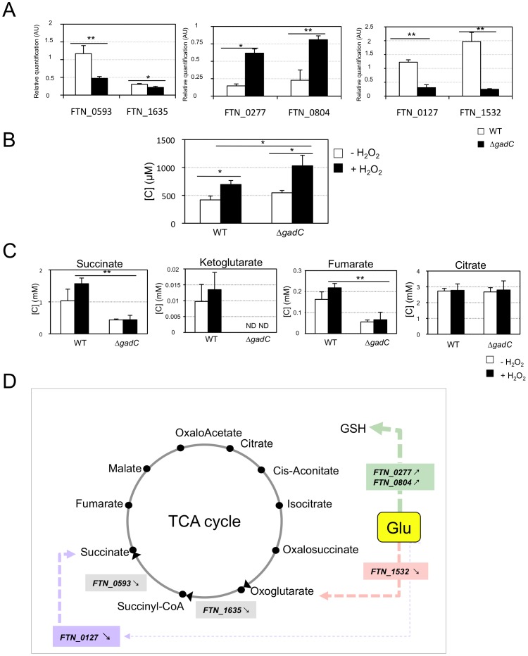
means the ratio (H2O2-treated/non-treated) is lower in the mutant strain than in the wild-type strain; ↗ the ratio (H2O2-treated/non-treated) is higher in the mutant strain than in the wild-type strain. In the absence of external glutamate (e.g. in standard chemically defined medium), the pool of glutamate present in the bacterial cytoplasm may be synthesized either from oxoglutarate, glutamine, GSH or even proline (according to KEGG metabolic pathways).

Similar articles

Cited by

References

    1. Sjostedt A, editor (2011) Francisella tularensis and tularemia: Fontiers Media SA. - PMC - PubMed
    1. Oyston PC, Sjostedt A, Titball RW (2004) Tularaemia: bioterrorism defence renews interest in Francisella tularensis . Nat Rev Microbiol 2: 967–978. - PubMed
    1. Keim P, Johansson A, Wagner DM (2007) Molecular epidemiolgy, evolution, and ecology of Francisella . Ann N Y Acad Sci 1105: 30–66. - PubMed
    1. Celli J, Zahrt TC (2013) Mechanisms of Francisella tularensis intracellular pathogenesis. Cold Spring Harb Perspect Med 3: a010314. - PMC - PubMed
    1. McCaffrey RL, Schwartz JT, Lindemann SR, Moreland JG, Buchan BW, et al. (2010) Multiple mechanisms of NADPH oxidase inhibition by type A and type B Francisella tularensis . J Leukoc Biol 88: 791–805. - PMC - PubMed

Publication types

MeSH terms

LinkOut - more resources