. 2014 Jan;10(1):e1004080.
doi: 10.1371/journal.pgen.1004080. Epub 2014 Jan 16.

Bioelectric signaling regulates size in zebrafish fins

Affiliations

Bioelectric signaling regulates size in zebrafish fins

Simon Perathoner et al. PLoS Genet. 2014 Jan.

Abstract

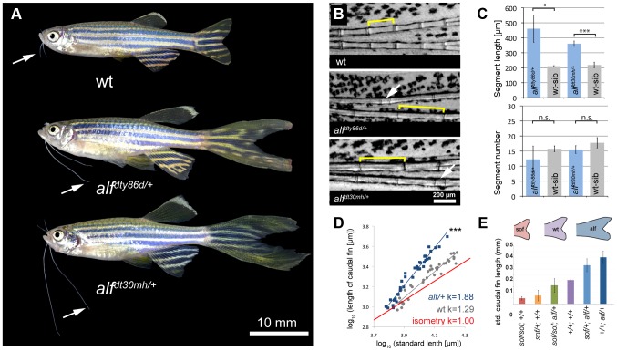
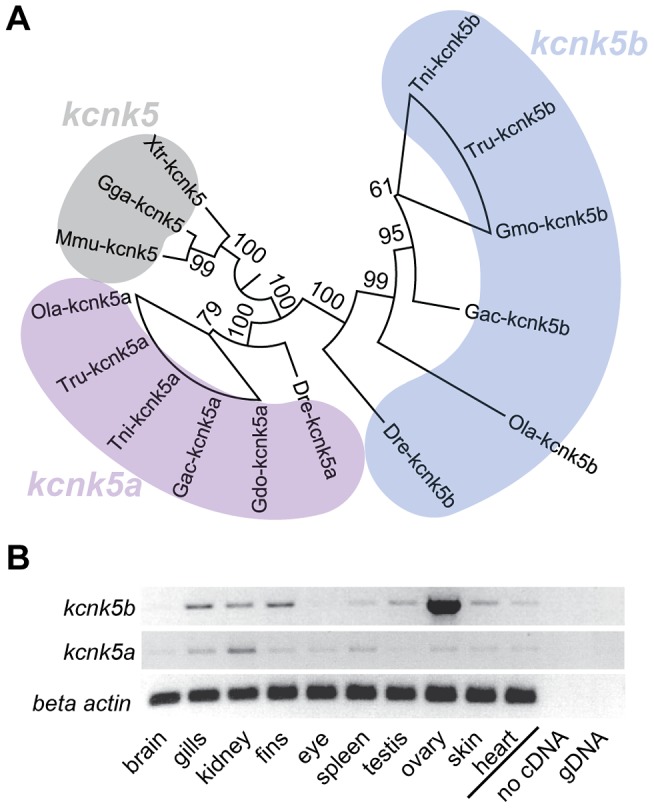
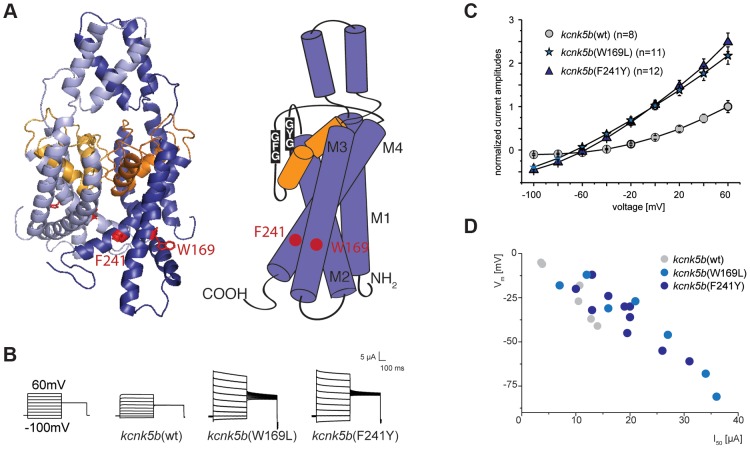
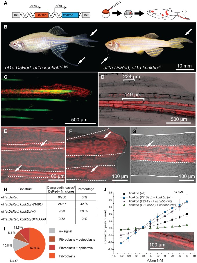
The scaling relationship between the size of an appendage or organ and that of the body as a whole is tightly regulated during animal development. If a structure grows at a different rate than the rest of the body, this process is termed allometric growth. The zebrafish another longfin (alf) mutant shows allometric growth resulting in proportionally enlarged fins and barbels. We took advantage of this mutant to study the regulation of size in vertebrates. Here, we show that alf mutants carry gain-of-function mutations in kcnk5b, a gene encoding a two-pore domain potassium (K(+)) channel. Electrophysiological analysis in Xenopus oocytes reveals that these mutations cause an increase in K(+) conductance of the channel and lead to hyperpolarization of the cell. Further, somatic transgenesis experiments indicate that kcnk5b acts locally within the mesenchyme of fins and barbels to specify appendage size. Finally, we show that the channel requires the ability to conduct K(+) ions to increase the size of these structures. Our results provide evidence for a role of bioelectric signaling through K(+) channels in the regulation of allometric scaling and coordination of growth in the zebrafish.


Conflict of interest statement

The authors have declared that no competing interests exist.

Figures

Figure 1
Figure 1. alf mutants lead to an increase in size of the appendages of adult fish.
(A) alf mutations are dominant and lead to overgrown fins and barbels in the adult. Arrows indicate maxillary barbels; the mutants shown are heterozygous. (B) Segment patterning in the dorsal fin of wild type and heterozygous mutants. Brackets indicate one segment. Although the majority of segments show increased length, several short segments can be seen in the mutants (arrows). (C) Variation in segment length (top) and segment number (bottom) in the longest ray of the dorsal fin of mutants and wild type siblings (wt sib). Fish of similar standard length (SL) (i.e. distance between snout and caudal peduncle) were compared; all cases n = 4; error bars: standard deviation; n.s.: not significant; *: p<0.02, ***: p<0.001. (D) Increased allometric scaling of heterozygous alf fins in development. k = allometric coefficient, Linear regression lines, wt R2 = 0.92; alf/+, R2 = 0.95; ***: p<0.001. (E) Crosses of sof with alf indicate that there is not epistatic interaction between the two genes. Fin length was normalized with SL.
Figure 2
Figure 2. Cell proliferation is increased in alf mutants.
(A) Sections of wild type and heterozygous alf fins. No significant difference in cell size is seen in the two groups. (B) Antibody staining against PCNA on paraffin sections of regenerating fins 4 days post amputation (dpa). Chart shows percentage of proliferating nuclei (PCNA) over total nuclei (Hoechst). N = 3–4 sections of 4 individual fish **: p-value<0.01.
Figure 3
Figure 3. The alf phenotype is due to gain-of-function mutations within the K+ channel kcnk5b.
(A) alf mutations map to chromosome 20 between z11841 and z21067. Gray: north markers; blue: south markers. (B) Electropherogram of kcnk5b at position 169 and 241 in mutants and wild type siblings. (C) The amino acids affected in the mutants are well conserved among vertebrates. (D) A revertant of alfdty86d (j131x8) shows wild type-sized fins. (E) kcnk5bj131x8 fish harbor an intragenic deletion in kcnk5b that is predicted to cause a truncated protein lacking three transmembrane (TM) domains.
Figure 4
Figure 4. Vertebrate kcnk5 homologs and expression in zebrafish development.
(A) Due to a whole genome duplication event, teleost fish have two kcnk5 paralogs that show early divergence. Numbers indicate bootstrap values in percentage (100 bootstrap replications). Nodes with a bootstrap value lower than 95 were collapsed. Dre, Danio rerio; Ola, Oryzias latipes; Gac, Gasterosteus aculeatus, Tru, Takifugu rubripes; Tni Tetraodon nigridoviridis, Gmo, Gadus morhua; Mmu, mus musculus; Gga, Gallus gallus; Xtr Xenopus tropicalis. (B) RT-PCR of kcnk5a and kcnk5b shows comparable expression between the two paralogs in multiple adult tissues, including fins.
Figure 5
Figure 5. Gain-of-function mutations in kcnk5b affect ionic conduction and lead to hyperpolarization of the cell.
(A) Location of the amino acids altered in kcnk5b gain-of-function mutants. Kcnk5b protein was modeled on human KCNK4 (K2p4.1). GFG and GYG domains represent the selectivity pore of the channel. (B) Voltage clamp recordings from Xenopus oocytes injected with cRNA of wild type and mutant kcnk5b. The membrane potential was clamped at a reference potential of −80 mV and then stepped to a test potential from +60 mV to −100 mV for 500 ms. The current that is applied in order to clamp the voltage to a certain value corresponds to the current passing through the plasma membrane. Representative electrophysiological traces are shown. (C) The mutant channels display increased conductance over wild type channels expressed at comparable levels. Error bars represent standard deviation. (D) Kcnk5b influences membrane potential (Vm) in oocytes. The mutant variants tend to hyperpolarize the cell (each point represents one oocyte).
Figure 6
Figure 6. Overexpression of kcnk5b is sufficient to cause fin overgrowth.
(A) Construct used to create kcnk5b-expressing clones via Tol2 transgenesis. (B) Individual fish expressing kcnk5b (W169L) (left) or kcnk5b (wt) (right) in mosaic clones display localized fin and barbel overgrowth. (C–F) Overgrowth is associated with DsRed expression (in red) within mesenchymal cells. (C) Calcein staining labels bone tissue (in green) of an overgrown fin (DsRed; kcnk5(W169L) expressing clone). (D) Mesenchymal clones are associated with increased segment length in the fin compared to non-overgrown DsRed negative regions. (E) Fibroblast-like cells appear as DsRed positive cells within the fin rays (dotted line) that surround DsRed negative vasculature (arrows in E and F) which extend along the actinotrichia (fibrils within dotted lines in F) towards the distal end of the fin. (G) Overgrown barbels show DsRed signal within the mesenchyme (area within dotted line) but not in the vasculature (arrow). (H) Number of clones associated with overgrowth in different kcnk5b variants. (I) Proportion of different cell types labeled in overgrown tissues. (J) Electrophysiological recordings of the non-conductive kcnk5b (GFGAAA) mutant in oocytes. Squares: kcnk5b (wt), purple stars: kcnk5b (F241Y)+kcnk5b (wt), blue circles: kcnk5b (W169L)+kcnk5b (wt), green triangles: + kcnk5b (GFGAAA)+kcnk5b (wt). Current was normalized to the measurement of wt current at 60 mV. Inset: DsRed+ fibroblasts in fish injected with the non-conductive construct do not lead to fin overgrowth.

Similar articles

Cited by

References

    1. Lui JC, Baron J (2011) Mechanisms limiting body growth in mammals. Endocr Rev 32: 422–440. - PMC - PubMed
    1. Huxley JS, Teissier G (1936) Terminology of relative growth. Nature 137: 780–781.
    1. Gould SJ (1966) Allometry and size in ontogeny and phylogeny. Biol Rev Camb Philos Soc 41: 587–640. - PubMed
    1. Metcalf D (1963) The autonomous behaviour of normal thymus grafts. Aust J Exp Biol Med Sci 41: SUPPL437–447. - PubMed
    1. Metcalf D (1964) Restricted growth capacity of multiple spleen grafts. Transplantation 2: 387–392. - PubMed

Publication types