Review
. 2014 Jan;10(1):e1004086.
doi: 10.1371/journal.pgen.1004086. Epub 2014 Jan 16.

Is non-homologous end-joining really an inherently error-prone process?

Affiliations
Review

Is non-homologous end-joining really an inherently error-prone process?

Mireille Bétermier et al. PLoS Genet. 2014 Jan.

Abstract

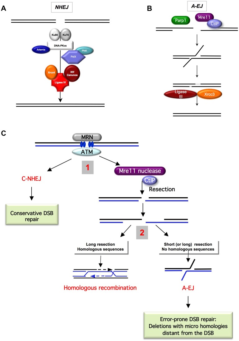
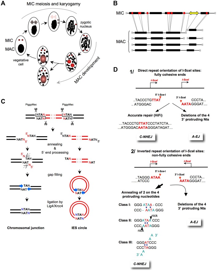
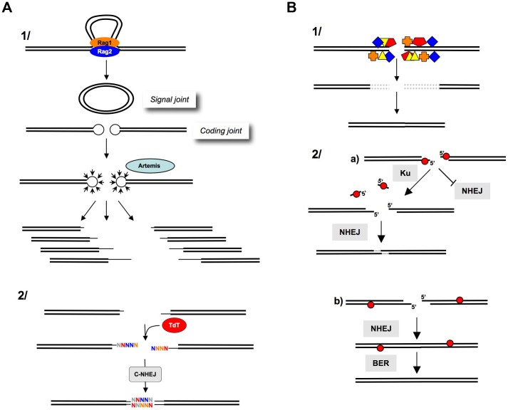
DNA double-strand breaks (DSBs) are harmful lesions leading to genomic instability or diversity. Non-homologous end-joining (NHEJ) is a prominent DSB repair pathway, which has long been considered to be error-prone. However, recent data have pointed to the intrinsic precision of NHEJ. Three reasons can account for the apparent fallibility of NHEJ: 1) the existence of a highly error-prone alternative end-joining process; 2) the adaptability of canonical C-NHEJ (Ku- and Xrcc4/ligase IV-dependent) to imperfect complementary ends; and 3) the requirement to first process chemically incompatible DNA ends that cannot be ligated directly. Thus, C-NHEJ is conservative but adaptable, and the accuracy of the repair is dictated by the structure of the DNA ends rather than by the C-NHEJ machinery. We present data from different organisms that describe the conservative/versatile properties of C-NHEJ. The advantages of the adaptability/versatility of C-NHEJ are discussed for the development of the immune repertoire and the resistance to ionizing radiation, especially at low doses, and for targeted genome manipulation.


Conflict of interest statement

The authors have declared that no competing interests exist.

Figures

Figure 1
Figure 1. End-joining models and competition between C-NHEJ and A-EJ for DSB repair.
A) The canonical C-NHEJ. The heterodimer Ku80-Ku70 binds to the DNA ends, which then recruit DNA-PKcs. Note that DNA-PK is absent from yeast. Several proteins, including Artemis, the polynucleotide kinase (PNK), and members of the polymerase X family, process the DNA ends for subsequent steps –. In the last step, ligase IV, associated with its co-factors Xrcc4 and Cernunos/Xlf, joins the ends –. B) A-EJ. Parp1 plays a role in the initiation process , , , . Without the protection by Ku70/Ku80, the DNA ends are resected in a reaction favored by the nuclease activity of Mre11 and CtIP , . It has been proposed that a single-strand DNA resection reveals complementary microhomologies (two to four nt or more) that can anneal; gap filling completes the end-joining. Subsequently, Xrcc1 and ligase III (which can be substituted by ligase I) complete A-EJ , , . A-EJ is always associated with deletions at the junctions and frequently (but not systematically) involves microhomologies that are distant from the DSB. The histone H1 has also been shown to act in A-EJ . C) Two-step model for the choice of the DSB repair pathway , . The MRN complex and ATM are involved in the early steps of DSB signaling and can activate both C-NHEJ and A-EJ. 1) Binding of Ku80/Ku70 protects from ssDNA resection, leading to a conservative DSB repair outcome through C-NHEJ. The nuclease activity of Mre11 and CtIP can initiate ssDNA resection. 2) A short ssDNA resection allows A-EJ but not homologous recombination. A long ssDNA resection allows A-EJ and HR, but HR requires the presence of homologous sequences. A-EJ results in error-prone repair associated with deletions at the repair junctions with frequent use of microhomologies distant from the DSB.
Figure 2
Figure 2. End-joining accuracy of ligation-compatible ends.
A) The Paramecium sexual cycle. Two types of sexual processes are induced through starvation in Paramecium: autogamy, a self-fertilization process (shown in the figure), and conjugation between compatible mating types (not shown). During autogamy, the two germline diploid MICs (red) undergo meiosis to generate eight haploid nuclei (pink), and a single nucleus migrates to a specialized cell compartment, dividing once to produce two identical gametic nuclei. The remaining seven meiotic products are degraded, and the old MAC (black) becomes fragmented. During karyogamy, the two gametic nuclei fuse to form a diploid zygotic nucleus. The zygotic nucleus subsequently undergoes two successive mitotic divisions; after the second division, the two nuclei become the new MICs of the sexual progeny (red), whereas the other two differentiate into new developing MACs (red and gray) and undergo programmed genomic rearrangements. At the first cell division, the new MICs divide mitotically, and each of the two developing new MACs segregates into a daughter cell where it continues to amplify the rearranged genome to a final ploidy of ∼800 n. During conjugation, MIC meiosis is triggered through the mating of two compatible sexual partners, which undergo a reciprocal exchange of their haploid gametic nuclei. Consequently, the zygotic nucleus in each partner is formed through the fusion of a resident and a migratory haploid nucleus. Exconjugants separate between the first and second divisions of the zygotic nucleus, and MAC development occurs as described for autogamous cells. B) General structure of MIC and MAC chromosomes. On the MIC chromosomes, genes (black boxes) and non-coding regions (thin lines) are interrupted by short internal eliminated sequences (IESs in red). Repeated germline sequences (e.g., transposons and minisatellites) are indicated with a yellow double-headed arrow. During MAC development, each MIC chromosome is amplified ∼400-fold to generate a population of heterogeneous MAC chromosomes. The imprecise elimination of repeated DNA is associated with the following alternative rearrangements: i) chromosome fragmentation and telomere addition to new MAC chromosome ends (gray squares) and ii) imprecise joining of the two chromosome arms that flank the eliminated germline region. C) Mechanism of IES excision. The successive DNA intermediates formed during IES excision are displayed, with IESs shown in red and flanking MAC-destined DNA shown in black. The first step of the reaction is the introduction of 4-base staggered double-strand breaks at each IES end, depending upon the PiggyMac domesticated transposase. The molecular steps that lead to the repair of the chromosomal junction are shown on the left, which might occur within a paired-end intermediate through the annealing of the central TAs within each 5′ overhang. The removal of the 5′-terminal nucleotide was demonstrated in vivo (dotted arrow), but the nuclease(s) involved has not been identified. For the 3′-processing step, ligase IV (Lig4) recruits or activates a gap-filling DNA polymerase, which adds one nucleotide to the recessive 3′-end prior to the final ligation. A similar mechanism has been proposed for the circularization of excised linear IES molecules (right), provided that these molecules are sufficiently long. IES circles do not replicate and are actively degraded. D) End-joining of fully versus non–fully complementary ends. 1) I- Sce I sites in direct orientation (arrows). The cleavage generates 3′-overhangs (red nt), which are fully complementary. C-NHEJ promotes accurate ligation (left panel), and A-EJ deletes the four protruding nucleotides, leading to the deletion of at least 4 bp at the resealed junction (right panel) , , . 2) I- Sce I sites in an inverted orientation (arrows). The cleavage generates 3′overhangs (red nt), which are not fully complementary. Similarly, A-EJ deletes the 3′-protruding nt, resulting in the deletion of at least 4 bp at the resealed junction (left panel). C-NHEJ anneals two of the four protruding nt (red nt), according to three classes of events (right panel). This imperfect annealing generates gaps (in blue in class I), mismatches (in blue in classes I and II), or 3′-single-stranded tails (in blue in class III) , , .
Figure 3
Figure 3. Processing of DNA ends prior to ligation.
A) Junctional diversity through V(D)J recombination. 1) The Rag1-Rag2 proteins join the V(D)J recombination sites (synapsis step). The cleavage by Rag1-Rag2 generates a circular signal joint and a linear coding joint (containing the coding sequence); however, the cleavage generates hairpins at the extremities, which cannot be directly ligated. The opening of the hairpins (by Artemis) generates a combination of different DNA ends (thin lines), thereby, creating the first level of junctional diversity. 2) TdT subsequently adds N-nucleotides at the 3′ or blunt ends, creating a second level of junctional diversity. B) After IR. 1) IR generates multiple damages at DNA ends (colored boxes). These altered DNA ends are not compatible for enzymatic ligation by ligase IV. The excision of the damaged structures (dotted lines) results in nucleotide deletion after ligation. 2) Role of Ku at the DNA ends. a) Abasic sites (red circles) at the DSB inhibit NHEJ. Ku removes these sites, allowing the NHEJ of the processed DNA ends. This reaction results in a limited deletion (one to three nt at the resealed junction). b) Abasic sites (red circles) that are far from the DSB do not impair NHEJ. BER can then repair the abasic sites on the resealed molecule. Note that the reduced activity of Ku on these substrates prevents long deletions (from the abasic site to the end), which would result in large deletions at the resealed junction and would avoid the generation of new breaks in the resealed molecule (adapted from [97]).

Similar articles

Cited by

References

    1. Guirouilh-Barbat J, Rass E, Plo I, Bertrand P, Lopez BS (2007) Defects in XRCC4 and KU80 differentially affect the joining of distal nonhomologous ends. Proc Natl Acad Sci U S A 104: 20902–20907. - PMC - PubMed
    1. Guirouilh-Barbat J, Huck S, Bertrand P, Pirzio L, Desmaze C, et al. (2004) Impact of the KU80 pathway on NHEJ-induced genome rearrangements in mammalian cells. Mol Cell 14: 611–623. - PubMed
    1. Grabarz A, Barascu A, Guirouilh-Barbat J, Lopez BS (2012) Initiation of DNA double strand break repair: signaling and single-stranded resection dictate the choice between homologous recombination, non-homologous end-joining and alternative end-joining. Am J Cancer Res 2: 249–268. - PMC - PubMed
    1. Audebert M, Salles B, Calsou P (2004) Involvement of poly(ADP-ribose) polymerase-1 and XRCC1/DNA ligase III in an alternative route for DNA double-strand breaks rejoining. J Biol Chem 279: 55117–55126. - PubMed
    1. Corneo B, Wendland RL, Deriano L, Cui X, Klein IA, et al. (2007) Rag mutations reveal robust alternative end joining. Nature 449: 483–486. - PubMed

Publication types

MeSH terms