. 2014 Jan;10(1):e1004107.
doi: 10.1371/journal.pgen.1004107. Epub 2014 Jan 16.

Cell-cycle dependent expression of a translocation-mediated fusion oncogene mediates checkpoint adaptation in rhabdomyosarcoma

Affiliations

Cell-cycle dependent expression of a translocation-mediated fusion oncogene mediates checkpoint adaptation in rhabdomyosarcoma

Ken Kikuchi et al. PLoS Genet. 2014 Jan.

Abstract

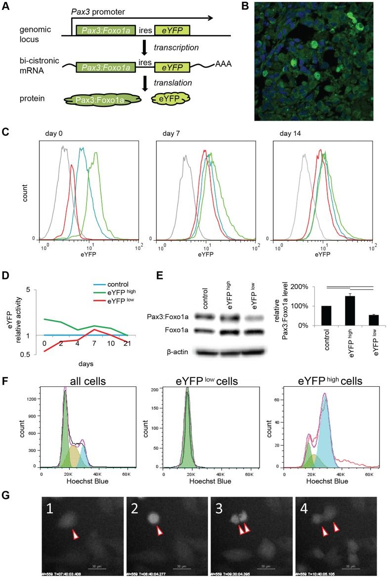
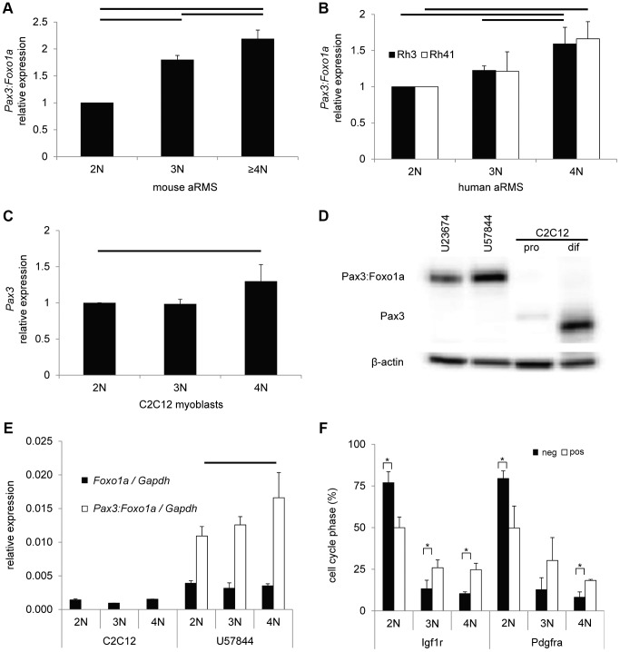
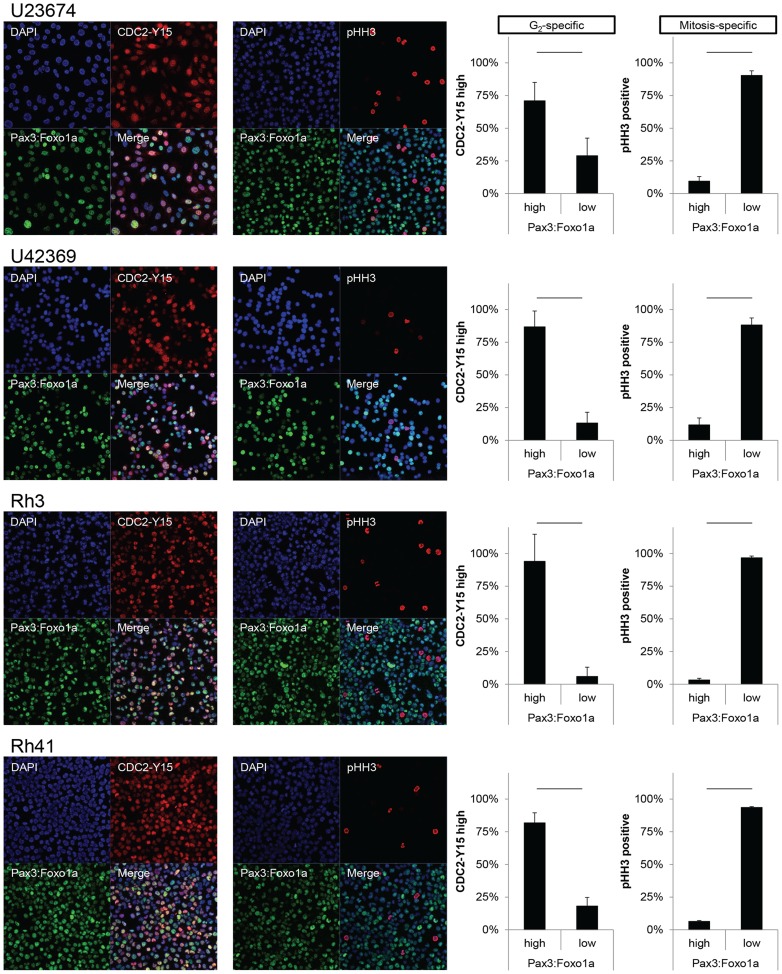
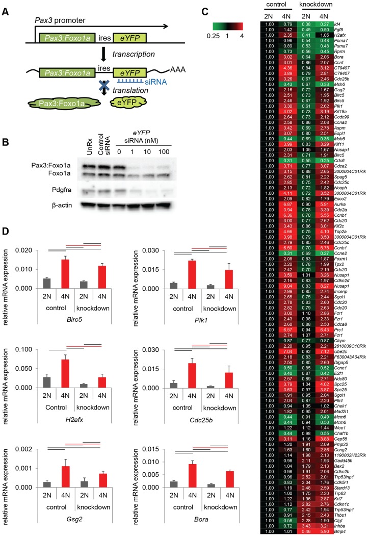
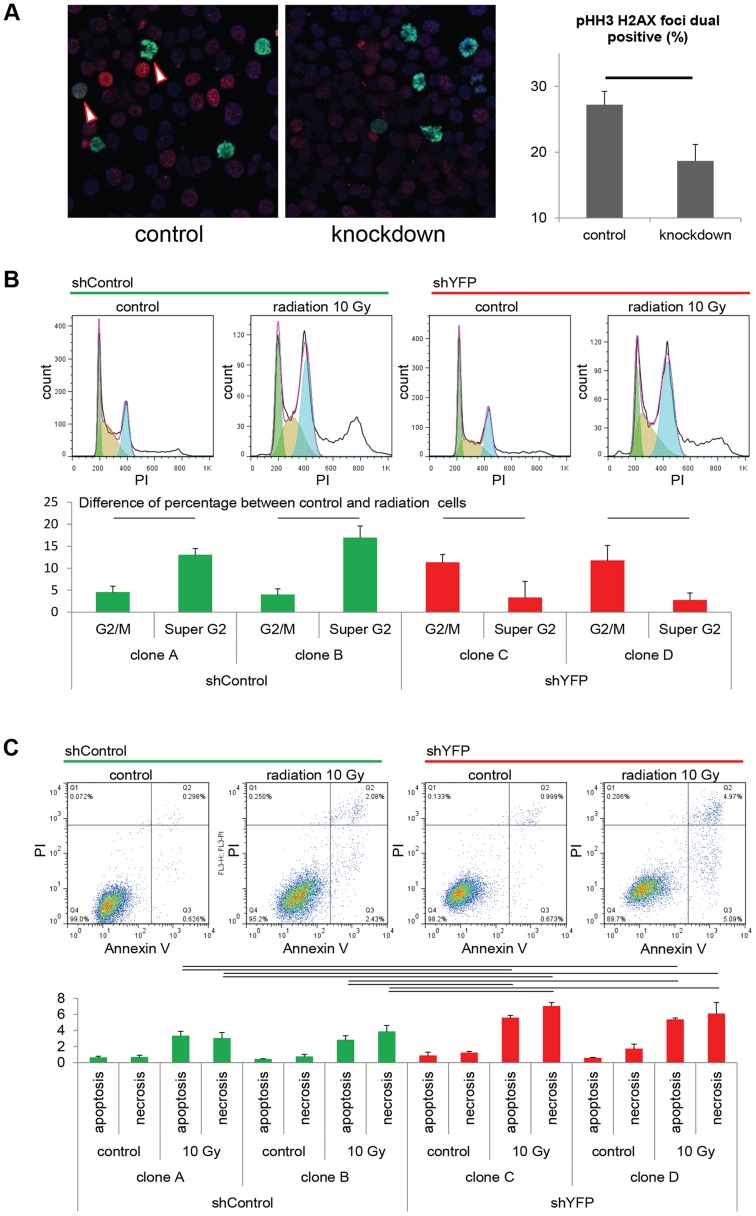
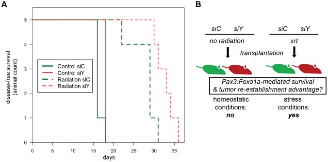
Rhabdomyosarcoma is the most commonly occurring soft-tissue sarcoma in childhood. Most rhabdomyosarcoma falls into one of two biologically distinct subgroups represented by alveolar or embryonal histology. The alveolar subtype harbors a translocation-mediated PAX3:FOXO1A fusion gene and has an extremely poor prognosis. However, tumor cells have heterogeneous expression for the fusion gene. Using a conditional genetic mouse model as well as human tumor cell lines, we show that that Pax3:Foxo1a expression is enriched in G2 and triggers a transcriptional program conducive to checkpoint adaptation under stress conditions such as irradiation in vitro and in vivo. Pax3:Foxo1a also tolerizes tumor cells to clinically-established chemotherapy agents and emerging molecularly-targeted agents. Thus, the surprisingly dynamic regulation of the Pax3:Foxo1a locus is a paradigm that has important implications for the way in which oncogenes are modeled in cancer cells.


Conflict of interest statement

The authors have declared that no competing interests exist.

Figures

Figure 1
Figure 1. eYFP activity and Pax3:Foxo1a expression is cell cycle specific.
(A) Diagrammatic representation of the conditional Pax3:Foxo1a knock-in allele by which eYFP is expressed as a second cistron on the same mRNA as Pax3:Foxo1a at the native Pax3 promoter. (B) Heterogeneity of eYFP expression in a murine aRMS tumor by immunofluorescence. (C) eYFP fluorescence of eYFP sorted cells overtime as measured by FACS. Grey: C2C12 (negative control), blue: no sorted cells, green: eYFP activity high cells, red: eYFP activity low cells. (D) Mean of relative eYFP activity in panel 1C measured by FACS. (E) Western blot analysis using eYFP sorted cells. Plotted are relative protein levels of Pax3:Foxo1a/β-Actin. Mean ± SE were obtained from three independent immunoblottings. Black line shows significant difference (p<0.05). (F) eYFP activity and cell cycle analysis using Hochest33342 staining for mouse primary cell culture U23674. Green shows G0/G1 phase, brown shows S phase, and blue shows G2/M phase. (G) Time-lapse experiment of eYFP activity (select frames over 16 hours). See also corresponding Movie S1.
Figure 2
Figure 2. Pax3:Foxo1a activity is cell cycle dependent.
(A) mRNA expression of Pax3:Foxo1a normalized by Gapdh in U23674 mouse aRMS primary cell culture sorted by DNA content. Black lines show significant differences (p<0.05). (B) mRNA expression of PAX3:FOXO1A normalized by GAPDH in Rh3 and Rh41 human aRMS cell lines sorted by DNA content. (C) mRNA expression of Pax3 normalized by Gapdh in C2C12 murine myoblasts sorted by DNA content. (D) Western blot analysis of Pax3 and Pax3:Foxo1a in unsorted murine U23674 aRMS cells (genotype Pax3 (Pax3:Foxo1a activated/Pax3:Foxo1a activated)), murine U57844 aRMS cells (genotype Pax3 (wt/Pax3:Foxo1a activated)), proliferative C2C12 myoblasts (pro) and differentiating C2C12 myoblasts (dif). (E) mRNA expression of Foxo1 and Pax3:Foxo1a normalized to Gapdh in C2C12 myoblasts and the U57844 mouse aRMS primary cell culture. (F) Cell cycle analysis after sorting for Pax3:Foxo1a targets Igf1r or Pdgfra in mouse aRMS tumor cells. Nearly twice as many 4N cells are Igf1r (or Pdgfra) positive versus Igf1r (or Pdgfra negative), suggesting these Pax3:Foxo1a targets may have a functional role late in the cell cycle (* P<0.05). pos, positive. Neg, negative.
Figure 3
Figure 3. Pax3:Foxo1a is expressed in G2 for mouse and human aRMS.
Immunocytochemistry for Pax3 (green), pHH3 (red) and DAPI (blue) or Pax3 (green), pCDC2 (red) and DAPI (blue). Numbers are relative rate of Pax3:Foxo1a high or low cells/pHH3 positive cells and Pax3:Foxo1a high or low cells/CDC2-Y15 high cells. Black line shows significant difference (p<0.05).
Figure 4
Figure 4. Pax3:Foxo1a induces G2/M checkpoint adaptation gene in G2/M.
(A) Diagrammatic representation of Pax3:Foxo1a knockdown strategy using eYFP siRNA. (B) Knockdown of the Pax3:Foxo1a protein by siYFP. Total cell lysates were isolated 48 h after transfection. Pax3:Foxo1a was detected with an antibody targeting the C-terminus of Foxo1a. (C) Differential expression of 60 of cell cycle genes (as annotated by Gene Ontology) for DNA content with or without Pax3:Foxo1a knockdown. (D) mRNA expression by QPCR of Plk1, Cdc25b, H2afx and Birc5 normalized to Gapdh in DNA content-sorted U23674 mouse aRMS primary tumor cells with or without Pax3:Foxo1a knockdown. Black and red line shows significant difference (p<0.05).
Figure 5
Figure 5. Pax3:Foxo1a facilitates G2/M checkpoint adaptation.
(A) Immunocytochemistry for pHH3 (green), pH2AX (red) and DAPI (Blue) using U23674 mouse aRMS primary cell culture with or without Pax3:Foxo1a knockdown treated with 6 Gy irradiation. Black line shows significant difference (p<0.05). See Figure S2A for representative single-channel ICC images corresponding to Figure 5A. Arrowheads indicate pHH3 and pH2AX double positive cells. (B) Representative cell cycle analysis for U23674 transfected by shControl (Clone A) or shYFP (Clone C) with or without 10 Gy irradiation. The graph shows the differences of percentage between control and radiated cells in shControl and shYFP clones in 3 independent experiments. Black line shows significant difference (p<0.05). (C) Annexin V apoptosis detection assay for U23674 transfected by shControl or shYFP clones with or without 10 Gy irradiation. Black line shows significant difference (p<0.05).
Figure 6
Figure 6. Treatment-related implications for dynamic oncogene expression in rhabdomyosarcoma in vivo.
(A) Kaplan-Meier survival analysis for disease-free survival of mice implanted with pre-irradiated (10Gy) primary murine aRMS tumor cells treated with Pax3:Foxo1a siRNA (siY) or control siRNA (siC), n = 5 animals per cohort. The p value for the difference between siY and siC groups receiving radiation was 0.02. (B) Diagrammatic representation of results in (A).

Comment in

References

    1. Breneman JC, Lyden E, Pappo AS, Link MP, Anderson JR, et al. (2003) Prognostic factors and clinical outcomes in children and adolescents with metastatic rhabdomyosarcoma–a report from the Intergroup Rhabdomyosarcoma Study IV. J Clin Oncol 21: 78–84. - PubMed
    1. Anderson JR, Barr FG, Hawkins DS, Parham DM, Skapek SX, et al. (2010) Fusion-negative alveolar rhabdomyosarcoma: modification of risk stratification is premature. J Clin Oncol 28: e587–588; author reply e589–590. - PubMed
    1. Williamson D, Missiaglia E, de Reynies A, Pierron G, Thuille B, et al. (2010) Fusion gene-negative alveolar rhabdomyosarcoma is clinically and molecularly indistinguishable from embryonal rhabdomyosarcoma. J Clin Oncol 28: 2151–2158. - PubMed
    1. Wexler LH, Ladanyi M (2010) Diagnosing alveolar rhabdomyosarcoma: morphology must be coupled with fusion confirmation. J Clin Oncol 28: 2126–2128. - PubMed
    1. Keller C, Arenkiel BR, Coffin CM, El-Bardeesy N, DePinho RA, et al. (2004) Alveolar rhabdomyosarcomas in conditional Pax3:Fkhr mice: cooperativity of Ink4a/ARF and Trp53 loss of function. Genes Dev 18: 2614–2626. - PMC - PubMed

Publication types

MeSH terms

Associated data