. 2014 Jan 14;9(1):e73402.
doi: 10.1371/journal.pone.0073402. eCollection 2014.

PDGF, NT-3 and IGF-2 in combination induced transdifferentiation of muscle-derived stem cells into Schwann cell-like cells

Affiliations

PDGF, NT-3 and IGF-2 in combination induced transdifferentiation of muscle-derived stem cells into Schwann cell-like cells

Yi Tang et al. PLoS One. .

Abstract

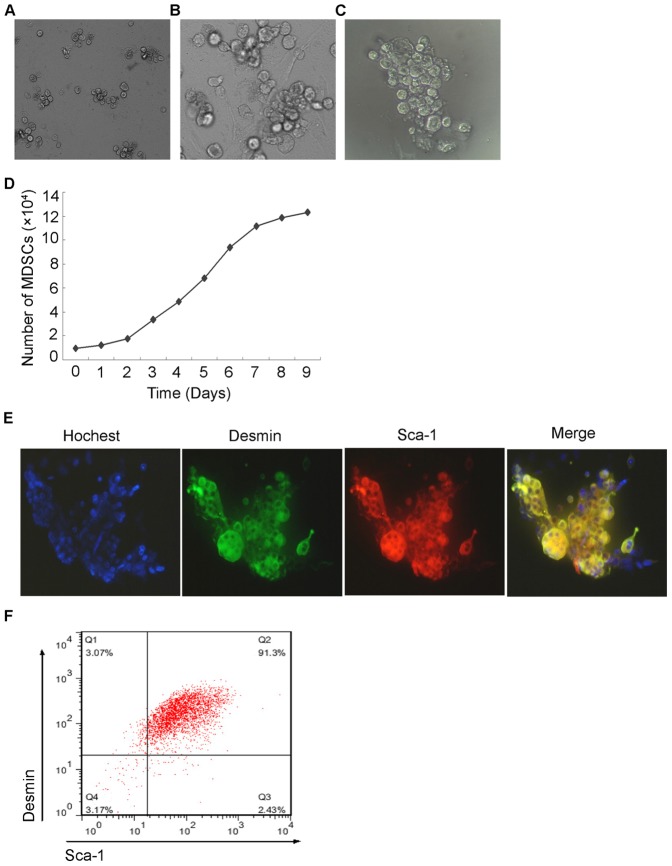
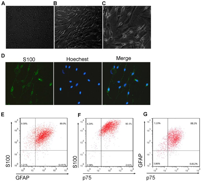
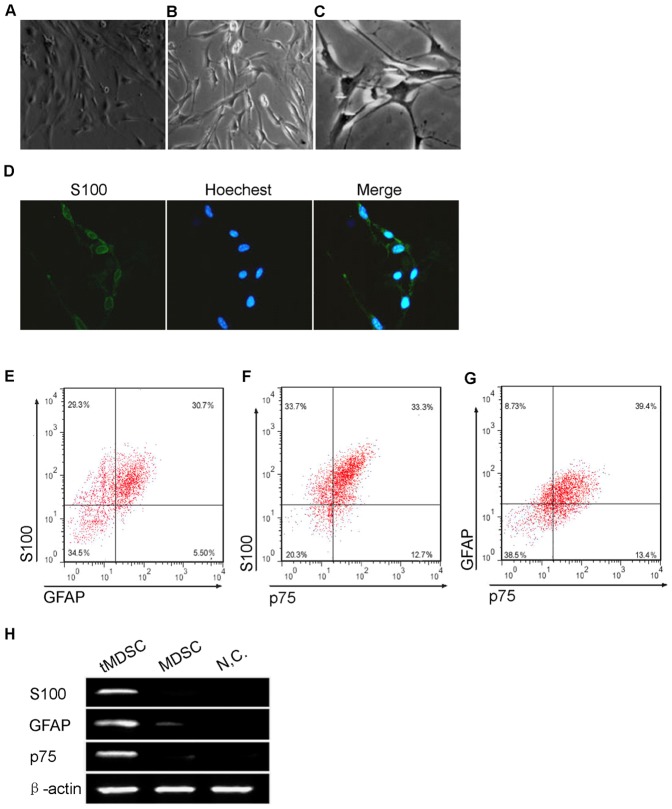
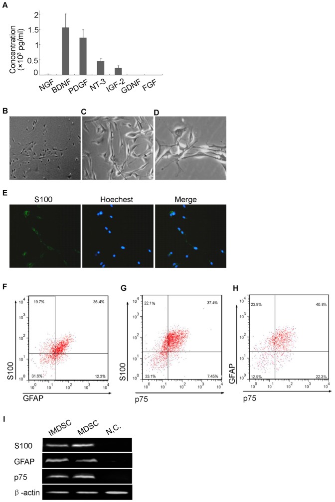
Muscle-derived stem cells (MDSCs) are multipotent stem cells with a remarkable long-term self-renewal and regeneration capacity. Here, we show that postnatal MDSCs could be transdifferentiated into Schwann cell-like cells upon the combined treatment of three neurotrophic factors (PDGF, NT-3 and IGF-2). The transdifferentiation of MDSCs was initially induced by Schwann cell (SC) conditioned medium. MDSCs adopted a spindle-like morphology similar to SCs after the transdifferentiation. Immunocytochemistry and immunoblot showed clearly that the SC markers S100, GFAP and p75 were expressed highly only after the transdifferentiation. Flow cytometry assay showed that the portion of S100 expressed cells was more than 60 percent and over one fourth of the transdifferentiated cells expressed all the three SC markers, indicating an efficient transdifferentiation. We then tested neurotrophic factors in the conditioned medium and found it was PDGF, NT-3 and IGF-2 in combination that conducted the transdifferentiation. Our findings demonstrate that it is possible to use specific neurotrophic factors to transdifferentiate MDSCs into Schwann cell-like cells, which might be therapeutically useful for clinical applications.


Conflict of interest statement

Competing Interests: The authors have declared that no competing interests exist.

Figures

Figure 1
Figure 1. Isolation and Characterisation of mouse MDSCs.
(A) Phase-contrast micrographs of undifferentiated PP6 cells isolated from mouse skeletal muscle. The cells had a rounded shape like stem cells. (B) Growth curve of PP6 cells showed a slow-cycling phenotype like stem cells. (C) Immunofluorescence staining showed the PP6 cells were desmin and Sca-1 positive which indicated they were MDSCs. (D) Flow cytometry assay demonstrated that 93.23±0.93% of the PP6 cells were Sca 1 positive, and 94.18±0.38% were desmin positive. 90.1±1.28% were double positive cells (data are mean % cells±S.E.M.). All experiments were repeated at least three times in triplicates.
Figure 2
Figure 2. Isolation and Characterisation of mouse SCs to obtain the conditioned medium.
(A) Phase-contrast micrographs of SCs isolated from mouse sciatic nerve and dorsal root ganglia. The cells had typical spindle-shaped SC morphology. (B) Immunofluorescence staining demonstrated the isolated cells expressed SC marker S100 protein. (C) Flow cytometry assay showed that most of the cells were S100 (96.77±1.46%), GFAP (92.92±4.94%), and p75 (93.38±0.90%) positive, and 86.12±1.53% of the cells expressed all of the three SC markers (data are mean % cells±S.E.M.).
Figure 3
Figure 3. Transdifferentiation of mouse MDSCs to a Schwann cell phenotype.
(A) Phase-contrast micrographs showed that MDSCs adopted a spindle-like shape with processes which was a characteristic of Schwann cells after treatment with SC conditioned medium. (B) Immunocytochemisty showed that the tMDSCs highly expressed the SC marker S100 protein. (C) Flow cytometry assay demonstrated that the portion of S100, GFAP, and p75 positive cells in tMDSCs were 65.48±6.20%, 39.84±1.66% and 41.08±0.78%, and 25.86±5.37% of the tMDSCs expressed all of the three SC markers (data are mean % cells±S.E.M.). (D) Immunoblot assay showed the MDSCs expressed of S100, GFAP and p75 only after transdifferentiation. NC, negative control. β-actin served as loading control. Right, bar graph of qualitative data in statistics. S100, GFAP and p75 protein levels were normalized to that of β-actin, shown as mean±S.E.M. **, p<0.01 vs. MDSC.
Figure 4
Figure 4. Neurotrophic factors which direct the transdifferentiation.
(A) ELISA assay showed the quantity of seven neurotrophic factors in the SC conditioned medium. There were high levels of BDNF, PDGF, NT-3 and IGF-2. The levels of NGF were relatively low, while GDNF and FGF were undetectable. (B) Phase-contrast micrographs showed that MDSCs adopted a SC-like shape upon treatment with PDGF (1000 pg/ml), NT-3 (500 pg/ml) and IGF-2 (200 pg/ml) in combination. (C) Immunocytochemisty showed that the tMDSCs highly expressed the SC marker S100 protein. (D) Flow cytometry assay demonstrated that the portion of S100, GFAP, and p75 positive cells in tMDSCs were 58.64±4.38%, 47.38±0.84% and 44.33±2.39%, while 27.89±5.98% of the tMDSCs expressed all of the three SC markers (data are mean % cells±S.E.M.). (E) Immunoblot assay showed the MDSCs expressed of S100, GFAP and p75 only after transdifferentiation. β-actin served as loading control. Right, bar graph of qualitative data in statistics. S100, GFAP and p75 protein levels were normalized to that of β-actin, shown as mean±S.E.M. **, p<0.01 vs. MDSC.

Similar articles

Cited by

References

    1. Bunge RP (1994) The role of the Schwann cell in trophic support and regeneration. J Neurol 242: 19–21. - PubMed
    1. Ide C (1996) Peripheral nerve regeneration. Neurosci Res 25: 101–21. - PubMed
    1. Jessen KR, Mirsky R (1999) Schwann cells and their precursors emerge as major regulators of nerve development. Trends Neurosci 22: 402–10. - PubMed
    1. Guenard V, Kleitman N, Morrissey TK, Bunge RP, Aebischer P (1992) Syngenic Schwann cells derived from adult nerves seeded in semi-permeable guidance channels enhance peripheral nerve regeneration. J Neurosci 12: 3310–20. - PMC - PubMed
    1. Mosahebi A, Woodward B, Wiberg M, Martin R, Terenghi G (2001) Retroviral labelling of Schwann cells: In vitro charactarisation and in vivo transplantation to improve peripheral nerve regeneration. Glia 34: 8–17. - PubMed

Publication types

MeSH terms

Substances