

Bat airway epithelial cells: a novel tool for the study of zoonotic viruses
- PMID: 24454736
- PMCID: PMC3890267
- DOI: 10.1371/journal.pone.0084679
Bat airway epithelial cells: a novel tool for the study of zoonotic viruses
Abstract
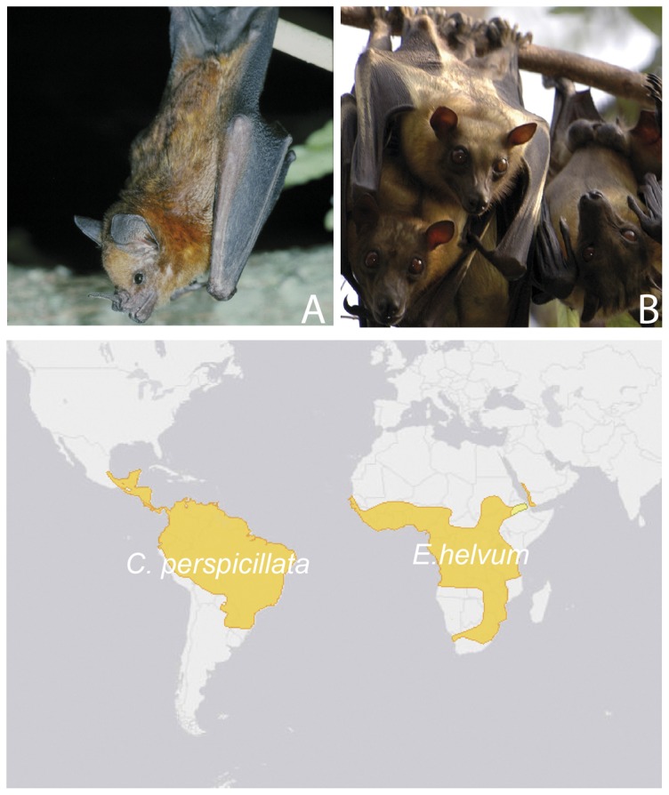
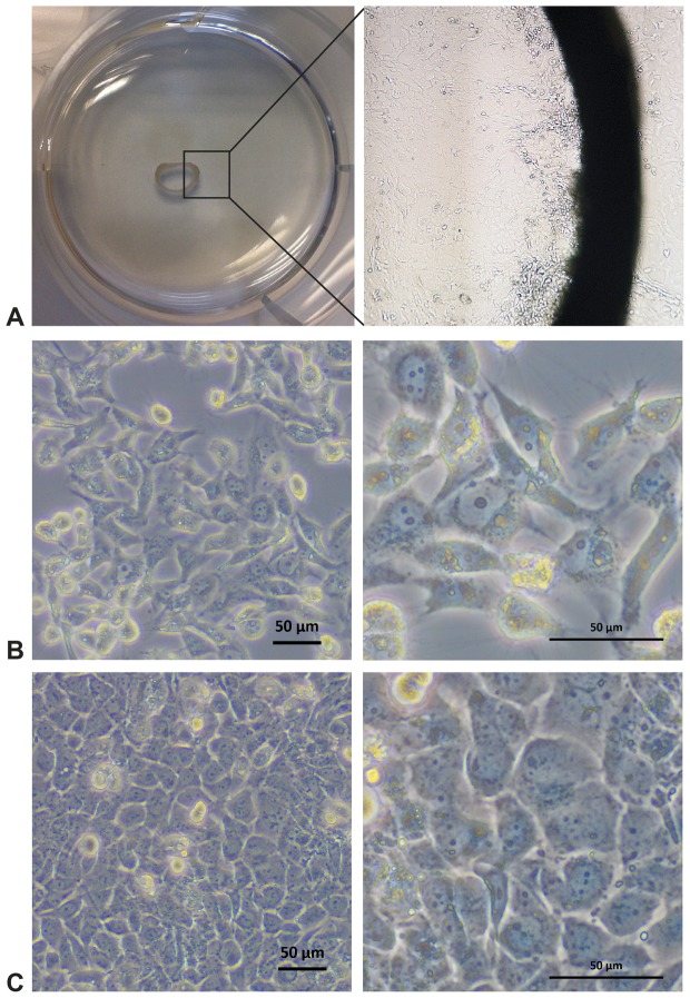
Bats have been increasingly recognized as reservoir of important zoonotic viruses. However, until now many attempts to isolate bat-borne viruses in cell culture have been unsuccessful. Further, experimental studies on reservoir host species have been limited by the difficulty of rearing these species. The epithelium of the respiratory tract plays a central role during airborne transmission, as it is the first tissue encountered by viral particles. Although several cell lines from bats were established recently, no well-characterized, selectively cultured airway epithelial cells were available so far. Here, primary cells and immortalized cell lines from bats of the two important suborders Yangochiroptera and Yinpterochiroptera, Carollia perspicillata (Seba's short-tailed bat) and Eidolon helvum (Straw-colored fruit bat), were successfully cultured under standardized conditions from both fresh and frozen organ specimens by cell outgrowth of organ explants and by the use of serum-free primary cell culture medium. Cells were immortalized to generate permanent cell lines. Cells were characterized for their epithelial properties such as expression of cytokeratin and tight junctions proteins and permissiveness for viral infection with Rift-Valley fever virus and vesicular stomatitis virus Indiana. These cells can serve as suitable models for the study of bat-borne viruses and complement cell culture models for virus infection in human airway epithelial cells.
Conflict of interest statement
Figures

Similar articles
-
Accelerated viral dynamics in bat cell lines, with implications for zoonotic emergence.Elife. 2020 Feb 3;9:e48401. doi: 10.7554/eLife.48401. Elife. 2020. PMID: 32011232 Free PMC article.
-
Characterization of Experimental Oro-Nasal Inoculation of Seba's Short-Tailed Bats (Carollia perspicillata) with Bat Influenza A Virus H18N11.Viruses. 2020 Feb 19;12(2):232. doi: 10.3390/v12020232. Viruses. 2020. PMID: 32093076 Free PMC article.
-
Infection Studies with Airway Organoids from Carollia perspicillata Indicate That the Respiratory Epithelium Is Not a Barrier for Interspecies Transmission of Influenza Viruses.Microbiol Spectr. 2023 Mar 14;11(2):e0309822. doi: 10.1128/spectrum.03098-22. Online ahead of print. Microbiol Spectr. 2023. PMID: 36916937 Free PMC article.
-
European bats as carriers of viruses with zoonotic potential.Viruses. 2014 Aug 13;6(8):3110-28. doi: 10.3390/v6083110. Viruses. 2014. PMID: 25123684 Free PMC article. Review.
-
Public health awareness of emerging zoonotic viruses of bats: a European perspective.Vector Borne Zoonotic Dis. 2006 Winter;6(4):315-24. doi: 10.1089/vbz.2006.6.315. Vector Borne Zoonotic Dis. 2006. PMID: 17187565 Review.
Cited by
-
Productive Propagation of Rift Valley Fever Phlebovirus Vaccine Strain MP-12 in Rousettus aegyptiacus Fruit Bats.Viruses. 2018 Nov 30;10(12):681. doi: 10.3390/v10120681. Viruses. 2018. PMID: 30513679 Free PMC article.
-
More novel hantaviruses and diversifying reservoir hosts--time for development of reservoir-derived cell culture models?Viruses. 2014 Feb 26;6(3):951-67. doi: 10.3390/v6030951. Viruses. 2014. PMID: 24576845 Free PMC article. Review.
-
Novel methods for in vitro modeling of pancreatic cancer reveal important aspects for successful primary cell culture.BMC Cancer. 2020 May 13;20(1):417. doi: 10.1186/s12885-020-06929-8. BMC Cancer. 2020. PMID: 32404074 Free PMC article.
-
Bats as instructive animal models for studying longevity and aging.Ann N Y Acad Sci. 2024 Nov;1541(1):10-23. doi: 10.1111/nyas.15233. Epub 2024 Oct 4. Ann N Y Acad Sci. 2024. PMID: 39365995 Free PMC article. Review.
-
Attenuation of replication by a 29 nucleotide deletion in SARS-coronavirus acquired during the early stages of human-to-human transmission.Sci Rep. 2018 Oct 11;8(1):15177. doi: 10.1038/s41598-018-33487-8. Sci Rep. 2018. PMID: 30310104 Free PMC article.
References
-
- Drosten C, Gunther S, Preiser W, van der Werf S, Brodt HR, et al. (2003) Identification of a novel coronavirus in patients with severe acute respiratory syndrome. N Engl J Med 348: 1967–1976. - PubMed
Publication types
MeSH terms
Substances
LinkOut - more resources
Full Text Sources
Other Literature Sources
Medical
Research Materials
