. 2014 Jan 14;9(1):e84845.
doi: 10.1371/journal.pone.0084845. eCollection 2014.

The effect of brain-derived neurotrophic factor on periodontal furcation defects

Affiliations

The effect of brain-derived neurotrophic factor on periodontal furcation defects

Ryo Jimbo et al. PLoS One. .

Abstract

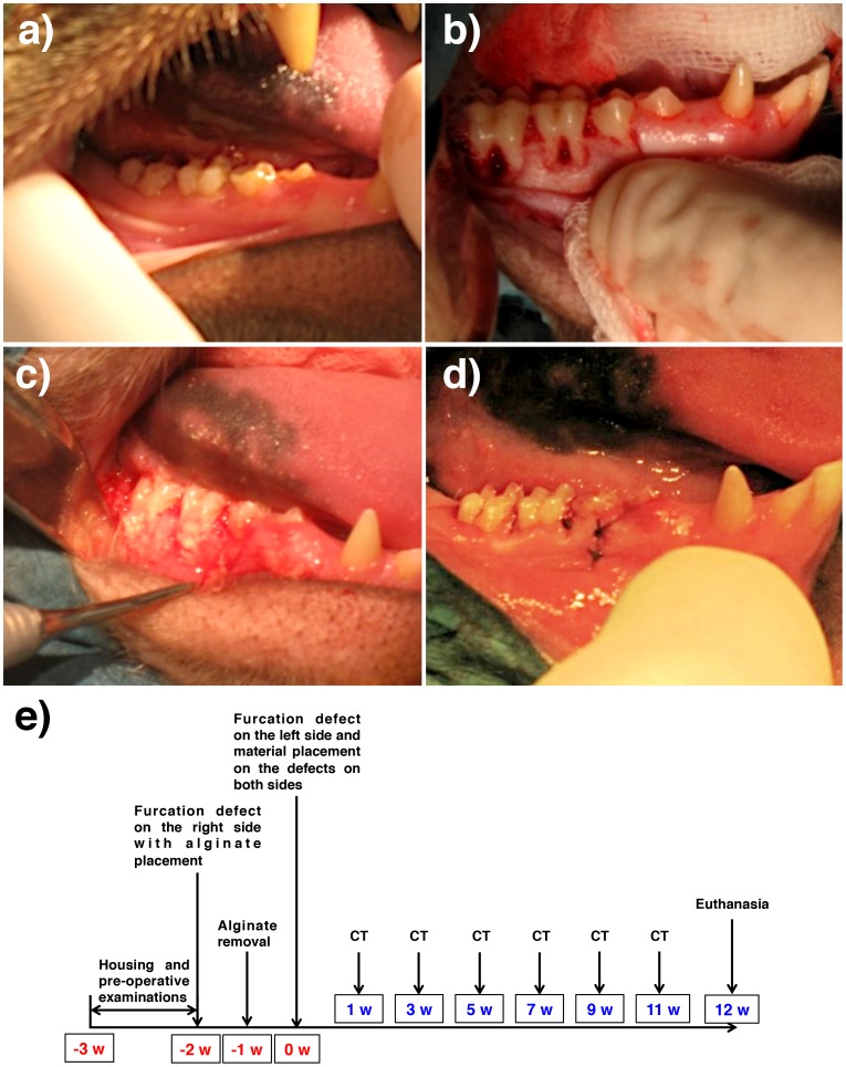
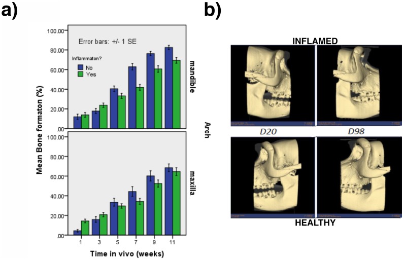
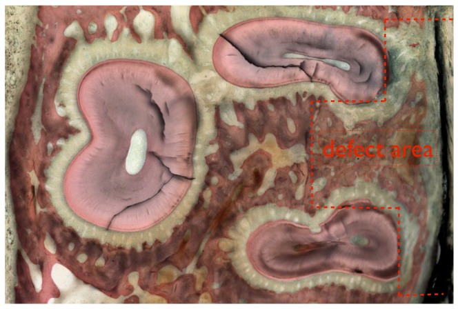
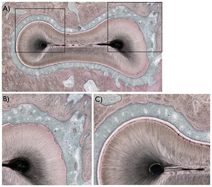
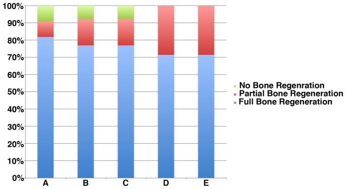
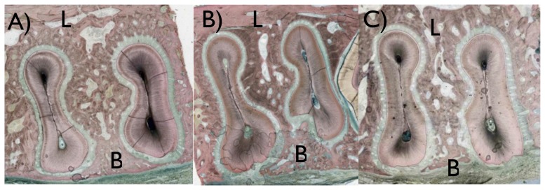
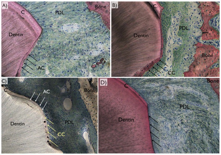
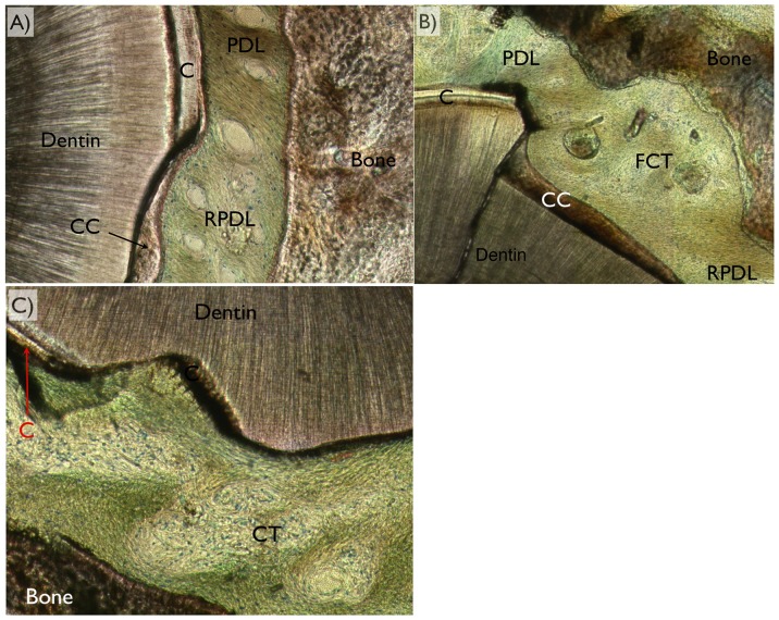
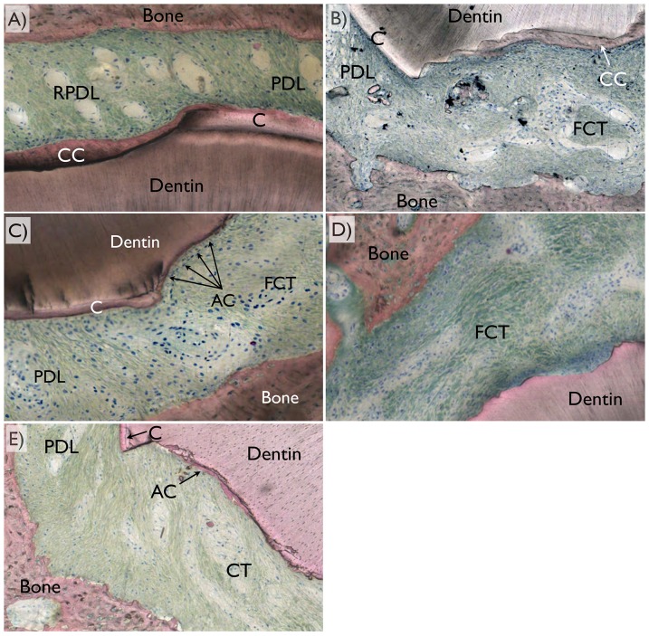
This study aimed to observe the regenerative effect of brain-derived neurotrophic factor (BDNF) in a non-human primate furcation defect model. Class II furcation defects were created in the first and second molars of 8 non-human primates to simulate a clinical situation. The defect was filled with either, Group A: BDNF (500 µg/ml) in high-molecular weight-hyaluronic acid (HMW-HA), Group B: BDNF (50 µg/ml) in HMW-HA, Group C: HMW-HA acid only, Group D: empty defect, or Group E: BDNF (500 µg/ml) in saline. The healing status for all groups was observed at different time-points with micro computed tomography. The animals were euthanized after 11 weeks, and the tooth-bone specimens were subjected to histologic processing. The results showed that all groups seemed to successfully regenerate the alveolar buccal bone, however, only Group A regenerated the entire periodontal tissue, i.e., alveolar bone, cementum and periodontal ligament. It is suggested that the use of BDNF in combination with a scaffold such as the hyaluronic acid in periodontal furcation defects may be an effective treatment option.


Conflict of interest statement

Competing Interests: This study was funded by Dentply IH, however, this does not alter the authors' adherence to all the PLOS ONE policies on sharing data and materials.

Figures

Figure 1
Figure 1. Design of the experiment.
(a) Pre operative condition of the oral cavity. All animals used were in periodontally healthy condition. (b) Full removal of the buccal plate and defect creation at the furcation region, (c) alginate placement to induce inflammation, and (d) suture. (e) The schematic timeline of the current study.
Figure 2
Figure 2. The comparison between induced inflammation versus non-inflamed sites.
(a) Graph representing the effect of interactions between Arch, Inflammation, and Time. (b) Representative 3D reconstructed image of the furcation defects healing overtime for both inflamed (top) and healthy (bottom) sites. No differences were seen in furcation defect healing for both conditions.
Figure 3
Figure 3. A descriptive histologic section showing the defect area (marked in red) for the maxillary molar.
Figure 4
Figure 4. Histologic images of the areas of interest observed in the study.
(a) A descriptive histologic section showing the region of interest evaluated (marked in black). (b) For all teeth evaluated, damage to the root was observed primarily at the buccal and furcation region of the roots, where regions of cementum were removed in full thickness and root dentin removed in partial thickness. (c) In regions not surgically affected, the typical anatomic features of hard (alveolar bone, cementum, and root dentin), and soft tissues (periodontal ligament) were observed.
Figure 5
Figure 5. Percent distribution of bone healing pattern, where majority of the samples presented full bone regeneration at the defect region, a smaller fraction partial regeneration, and an even smaller fraction of all samples of the study did not present full buccal bone regeneration.
Figure 6
Figure 6. Histologic sections showing the buccal (B) and lingual (L) regions of samples presenting different bone healing patterns.
Most of the sites evaluated presented (a) full bone regeneration, and few presented (b) partial regeneration or (c) no regeneration.
Figure 7
Figure 7. Representative histologic images depicting the periodontal ligament (PDL) region between bone and the tooth, where original cementum thickness (C) could be depicted in the vicinity of a defect.
The three different regeneration patterns comprised (a) acellular cementum (AC), (b) cellular cementum (CC), (c) mixed cellular and acellular cementum (MCA), or (d) no cementum regeneration (arrows).
Figure 8
Figure 8. For periodontal ligament regeneration assessment, optical microscopy in circular polarized mode was utilized and the different tissues (dentin, bone, cementum [C], cellular cementum [CC], acellular cementum [AC], original periodontal ligament [PDL], regenerated periodontal ligament [RPDL], and fibrous connective tissue [FCT were easily depicted).
Among the three variations of healing pattern observed were (a) full regeneration, (b) partial regeneration, and (c) no regeneration (no fibrous bridging between bone and cementum along the defect perimeter) were observed.
Figure 9
Figure 9. Representative histologic images depicting frequent healing patterns for each group depicting the region comprising bone, periodontal ligament region (PDL), and cementum (C).
(a) Group A primarily presented full bone and full defect perimeter cementum regeneration (cellular [CC] and full thickness repair), along with full periodontal ligament regeneration (RPDL) with fibers bridging the gap between the cellular cementum and newly formed bone. (b) Group B primarily presented primarily full bone regeneration along with partial cementum coverage (mostly celular at covered regions) and partial periodontal ligament regeneration. (c) Group C presented full bone and cementum regeneration (partial thickness, acellular) along with partial periodontal ligament regeneration. (d) Group D presented full bone regeneration with partial (mostly acellular) or no cementum regeneration, along with partial or no periodontal ligament regeneration. (e) Group E presented full bone regeneration with partial (mostly acellular) or no cementum regeneration, along with partial or no periodontal ligament regeneration.

Similar articles

Cited by

References

    1. Lindsay RM, Thoenen H, Barde YA (1985) Placode and neural crest-derived sensory neurons are responsive at early developmental stages to brain-derived neurotrophic factor. Dev Biol 112: 319–328. - PubMed
    1. Kubo T, Nonomura T, Enokido Y, Hatanaka H (1995) Brain-derived neurotrophic factor (BDNF) can prevent apoptosis of rat cerebellar granule neurons in culture. Brain Res Dev Brain Res 85: 249–258. - PubMed
    1. Pang PT, Teng HK, Zaitsev E, Woo NT, Sakata K, et al. (2004) Cleavage of proBDNF by tPA/plasmin is essential for long-term hippocampal plasticity. Science 306: 487–491. - PubMed
    1. Bekinschtein P, Cammarota M, Katche C, Slipczuk L, Rossato JI, et al. (2008) BDNF is essential to promote persistence of long-term memory storage. Proc Natl Acad Sci U S A 105: 2711–2716. - PMC - PubMed
    1. Nagahara AH, Tuszynski MH (2011) Potential therapeutic uses of BDNF in neurological and psychiatric disorders. Nat Rev Drug Discov 10: 209–219. - PubMed

Publication types

MeSH terms

Substances

LinkOut - more resources