Comparative Study
. 2014 Jan 15;9(1):e84973.
doi: 10.1371/journal.pone.0084973. eCollection 2014.

Comparative structural and functional analysis of orthomyxovirus polymerase cap-snatching domains

Affiliations
Comparative Study

Comparative structural and functional analysis of orthomyxovirus polymerase cap-snatching domains

Delphine Guilligay et al. PLoS One. .

Abstract

Orthomyxovirus Influenza A virus (IAV) heterotrimeric polymerase performs transcription of viral mRNAs by cap-snatching, which involves generation of capped primers by host pre-mRNA binding via the PB2 subunit cap-binding site and cleavage 10-13 nucleotides from the 5' cap by the PA subunit endonuclease. Thogotoviruses, tick-borne orthomyxoviruses that includes Thogoto (THOV), Dhori (DHOV) and Jos (JOSV) viruses, are thought to perform cap-snatching by cleaving directly after the cap and thus have no heterogeneous, host-derived sequences at the 5' extremity of their mRNAs. Based on recent work identifying the cap-binding and endonuclease domains in IAV polymerase, we determined the crystal structures of two THOV PB2 domains, the putative cap-binding and the so-called '627-domain', and the structures of the putative endonuclease domains (PA-Nter) of THOV and DHOV. Despite low sequence similarity, corresponding domains have the same fold confirming the overall architectural similarity of orthomyxovirus polymerases. However the putative Thogotovirus cap-snatching domains in PA and PB2 have non-conservative substitutions of key active site residues. Biochemical analysis confirms that, unlike the IAV domains, the THOV and DHOV PA-Nter domains do not bind divalent cations and have no endonuclease activity and the THOV central PB2 domain does not bind cap analogues. On the other hand, sequence analysis suggests that other, non-influenza, orthomyxoviruses, such as salmon anemia virus (isavirus) and Quaranfil virus likely conserve active cap-snatching domains correlating with the reported occurrence of heterogeneous, host-derived sequences at the 5' end of the mRNAs of these viruses. These results highlight the unusual nature of transcription initiation by Thogotoviruses.


Conflict of interest statement

Competing Interests: The authors have declared that no competing interests exist.

Figures

Figure 1
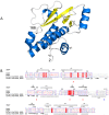
Figure 1. Structure of THOV PA-Nter domain.
A. Ribbon diagram showing the structure of the THOV PA-Nter domain. Helices are in cyan and beta strands in yellow and labeled according to the alignment in (B). B. Structure based sequence alignment of THOV, DHOV and IAV PA-Nter domains showing secondary structure elements for THOV and INFA. Red triangles indicate cation binding residues in IAV (His41, Glu80, Asp108 and Glu119). A blue triangle indicates the catalytic lysine (Lys134) in IAV which is on helix αA in INFA that does not exist in THOV and DHOV.
Figure 2
Figure 2. Structural comparison of THOV, DHOV and IAV PA-Nter domains.
A. Comparative structures of THOV, DHOV and IAV PA-Nter domains, in the same orientation after superposition, with coloring and secondary structure elements as in Figure 1A. Note helix αA (arrowed in right panel), which carries the catalytic lysine in IAV, is replaced by an irregular strand in THOV and DHOV. The root-mean-square deviation (RMSD) between THOV and DHOV is 1.47 Å for 142/169 aligned Cα<3.8 Å apart, and between THOV and IAV is 1.62 Å for 122 aligned Cα<3.8 Å apart. B. Electrostatic surfaces for the three domains (red, negatively charged; blue, positively charged). The IAV active site (right, arrowed) is negatively charged with a rim of positive charge, whereas THOV and DHOV have more positively charged residues within the active site. C. Comparison of residues in THOV and DHOV equivalent to the functionally important active site residues of IAV including the two bound cations. Only Asp86, Asp87 and Asp108 are conserved in THOV, DHOV and IAV, respectively.
Figure 3
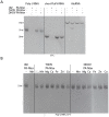
Figure 3. Functional comparison of THOV, DHOV and IAV PA-Nter domains.
A. Denaturing urea gel analysis of an endonuclease assay in which IAV, DHOV and THOV PA-Nter domains were incubated at 37°C for 30 minutes in the presence of 1 mM MnCl2 with U-rich RNA (left lanes), short Flu panhandle RNA (middle lanes) or Alu domain RNA (right lanes) as described . IAV endonuclease readily degrades the ssRNA and partially the structured Alu domain RNA, but THOV and DHOV PA-Nter are inactive under these conditions. B. Similar assay with THOV and DHOV PA-Nter and U-rich RNA in the presence or absence of various divalent ions (1 mM MnCl2, MgCl2, CaCl2, FeCl2, ZnCl2 or CoCl2). IAV endonuclease degrades the RNA in the presence of manganese but THOV PA-Nter does not degrade the RNA with any cation.
Figure 4
Figure 4. Structural comparison of THOV and IAV central PB2 domains.
A. Ribbon diagram comparing the structure of the IAV (left) and THOV (right) central PB2 domains, in the same orientation after superposition. Helices are in cyan and beta strands in yellow and labeled according to the alignment in (B). In the IAV, the bound cap analogue m7GTP is depicted together with three aromatic residues (Phe323, His357 and Phe404) involved in ligand binding. In the THOV domain, Arg344, which occupies the position equivalent to that of the m7G base, and Tyr413 (equivalent to Phe404) are shown. Comparison of the THOV and IAV domains using PDBeFOLD (http://www.ebi.ac.uk/msd-srv/ssm/) gives an RMSD = 3.0 Å for 133/160 matched Cα and Z = 6.1. B. Structure based sequence alignment of THOV and IAV central PB2 domains showing secondary structure elements. Blue triangles indicate the key residues involved in ligand binding in the case of IAV (Phe323, His357, Glu361, Phe363, Lys376 and Phe404). Red triangles indicate THOV residues that would clash with bound m7GTP (Met328 and Arg344, see Figure 5A).
Figure 5
Figure 5. Functional comparison of THOV and IAV central PB2 domains.
A. Comparison of m7GTP binding site and key interacting residues in IAV (left) and structurally equivalent residues in THOV (right) central PB2 domain. Colours as in (Figure 4A). Guanine base specific contacting residues in IAV (Glu361 and Lys376) are non-conservatively substituted by Ala370 and Cys385 in THOV, respectively and the aromatic residue Phe366 is differently orientated in THOV from His357 in IAV. In the THOV domain Arg344 occupies the position equivalent to that of the m7G base and Met328 that of the ribose and only aromatic residue Tyr413 (equivalent to Phe404 in IAV) is conservatively substituted. B. Cap-binding assay with THOV and IAV central PB2 domains. SDS PAGE analysis of the results of elution of IAV (IAV-capBD) and putative THOV cap binding domains (THOV-capBD) after binding on 7-methyl-GTP sepharose resin at 4°C. IAV-capBD (left lanes) was used as a positive control and binds the resin, whereas THOV-capBD (right lanes) does not. (IN) and (E) indicate input and eluted fractions respectively. Molecular weight markers (MW, kDa)) are shown in the right most lane. C. Minireplicon assay of the PB2 R344A mutant. THOV polymerase activity was determined in a reconstituted minireplicon system by transfecting 293T cells for 36 h with expression plasmids coding for a FF-Luc-encoding viral minigenome and viral proteins PB2, PB1, PA and NP. The PB2 subunit was transfected as Flag-tagged PB2(wt)(250 ng) or PB2(R344A)(350 ng) constructs to allow detection of the recombinant proteins by Western blot using specific antibodies. Firefly luciferase activities were normalized to Renilla. Mean values from two independent experiments with duplicates are shown. The error bars represent standard deviation.
Figure 6
Figure 6. Structural comparison of THOV and IAV PB2 627-domain.
A. Ribbon diagram comparing the structure of the IAV (left) and THOV (right) PB2 627 domains, in the same orientation after superposition. Helices are in cyan and beta strands in yellow and labeled according to the alignment in (B). The species specific residue Lys627 is shown for IAV; the corresponding loop in THOV is two residues longer. Comparison of the THOV and IAV (PDB 2VY7) domains using PDBeFOLD gives an RMSD = 2.6 Å for 114/154 matched Cα and Z = 5.0. B. Structure based sequence alignment of THOV and IAV PB2 627 domains showing secondary structure elements. Lys627 in the IAV domain is highlight with a green asterisk.

Similar articles

Cited by

References

    1. Kawaoka Y, Palese P (2005) Family Orthomyxoviridae. In: Fauquet CM, Mayo MA, Maniloff J, Desselberger U, Ball L, editors. Virus Taxonomy: Eighth Report of the International Committee on Taxonomy of Viruses,. San Diego: Elsevier Academic Press. pp. 681–693.
    1. Presti RM, Zhao G, Beatty WL, Mihindukulasuriya KA, da Rosa AP, et al. (2009) Quaranfil, Johnston Atoll, and Lake Chad viruses are novel members of the family Orthomyxoviridae. J Virol 83: 11599–11606. - PMC - PubMed
    1. Haig DA, Woodall JP, Danskin D (1965) Thogoto Virus: A Hitherto Underscribed Agent Isolated from Ticks in Kenya. Journal of general microbiology 38: 389–394. - PubMed
    1. Anderson CR, Casals J (1973) Dhori virus, a new agent isolated from Hyalomma dromedarii in India. The Indian journal of medical research 61: 1416–1420. - PubMed
    1. Moore DL, Causey OR, Carey DE, Reddy S, Cooke AR, et al. (1975) Arthropod-borne viral infections of man in Nigeria, 1964–1970. Ann Trop Med Parasitol 69: 49–64. - PubMed

Publication types

MeSH terms

LinkOut - more resources