. 2014 Jan 15;9(1):e85125.
doi: 10.1371/journal.pone.0085125. eCollection 2014.

Background synaptic activity in rat entorhinal cortex shows a progressively greater dominance of inhibition over excitation from deep to superficial layers

Affiliations

Background synaptic activity in rat entorhinal cortex shows a progressively greater dominance of inhibition over excitation from deep to superficial layers

Stuart David Greenhill et al. PLoS One. .

Abstract

The entorhinal cortex (EC) controls hippocampal input and output, playing major roles in memory and spatial navigation. Different layers of the EC subserve different functions and a number of studies have compared properties of neurones across layers. We have studied synaptic inhibition and excitation in EC neurones, and we have previously compared spontaneous synaptic release of glutamate and GABA using patch clamp recordings of synaptic currents in principal neurones of layers II (L2) and V (L5). Here, we add comparative studies in layer III (L3). Such studies essentially look at neuronal activity from a presynaptic viewpoint. To correlate this with the postsynaptic consequences of spontaneous transmitter release, we have determined global postsynaptic conductances mediated by the two transmitters, using a method to estimate conductances from membrane potential fluctuations. We have previously presented some of this data for L3 and now extend to L2 and L5. Inhibition dominates excitation in all layers but the ratio follows a clear rank order (highest to lowest) of L2>L3>L5. The variance of the background conductances was markedly higher for excitation and inhibition in L2 compared to L3 or L5. We also show that induction of synchronized network epileptiform activity by blockade of GABA inhibition reveals a relative reluctance of L2 to participate in such activity. This was associated with maintenance of a dominant background inhibition in L2, whereas in L3 and L5 the absolute level of inhibition fell below that of excitation, coincident with the appearance of synchronized discharges. Further experiments identified potential roles for competition for bicuculline by ambient GABA at the GABAA receptor, and strychnine-sensitive glycine receptors in residual inhibition in L2. We discuss our results in terms of control of excitability in neuronal subpopulations of EC neurones and what these may suggest for their functional roles.


Conflict of interest statement

Competing Interests: The authors have declared that no competing interests exist.

Figures

Figure 1
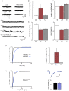
Figure 1. Characteristics of EPSCs in L3.
A. Voltage clamp recordings from one neurone show that the AMPAr antagonist NBQX essentially abolished sEPSCs. At more positive holding potentials, occasional, slow sEPSCs were detected that were mediated by NMDAr, as they were blocked by 2-AP5. B. A comparison of sEPSC properties Showed that the mean frequency of events in L3 was greater than that in either L2 or L5. Amplitudes were similar across layers, but kinetics (C) were slower in L2 compared to the deeper layers. D. Action potential driven release accounts for a much higher proportion of spontaneous excitation in L3. Cumulative probability analysis of data pooled from 10 neurones (graph on left) showed a large increase in IEI (shift to the right) in L3 that was substantially greater than that seen in L2 or L5. This is very clearly illustrated by the bar graphs (right) showing the accompanying change in frequencies (sEPSC frequency minus mEPSC frequency). E. Neither amplitude nor kinetics of EPSCs in the same neurones were significantly different in the presence of TTX (mEPSCs shown in blue).
Figure 2
Figure 2. Characteristics of IPSCs in L3.
A voltage clamp recordings from one neurone show that sIPSCs are largely mediated by GABAAr. However, there was a reduction in frequency with the glycine receptor antagonist strychnine. Although this generally only amounted to around 10–15% it was significant when assessed by a paired t-test. A similar effect was seen previously in L2 . We have added a number of new recordings to this data and the reduction again just reaches significance with a paired t-test. In contrast, no effect of strychnine was seen in L5. B. sIPSC frequency was lower than in L2 but still considerably higher than in L5, whereas the both mean amplitude and decay time in L3 (C) were greater than either of the other layers (D). Action potential driven GABA release, like glutamate release (see Figure 1) was more prominent in L3 compared to L2 or L5. Thus application of TTX resulted in a marked change in frequency of events (sEPSC frequency minus mIPSC frequency). (E) In addition, the mean amplitude of events was also lower in TTX, although time to decay was slower.
Figure 3
Figure 3. Comparison of inhibition to excitation in whole cell patch clamp studies.
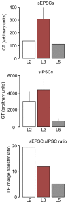
The total charge transfer (CT) associated with sEPSCs, was greatest in L3 and approximately equal in L2 and L5. Charge transfer of sIPSCs was also greatest in the middle layer, but substantially markedly less in L5 than the more superficial layers. The resultant ratio calculations suggested a substantial dominance of inhibition in all layers, with L2 showing by far the greatest bias and L5 the least.
Figure 4
Figure 4. Simultaneous VmD estimations of excitatory and inhibitory background conductances.
A. The raw traces show sample intracellular recordings from three neurones recorded in the same slice, and showing a great peak-to-peak voltage activity in L2. Estimation of background inhibition and excitation (see methods) required calculation of mean and variance of membrane potential at two levels of injected current (not shown). The histograms show the frequency distributions of membrane fluctuations at the two levels of injected current (geen being closer to resting potential and blue slightly more depolarized) in sample neurones from each layer. The solid lines on the graphs show the potential fluctuations constrained to a Gaussian function and in each case the goodness of fit (R2) was close to unity. B. Comparison of average data for background synaptic conductances estimated in the three layers of the EC. Background excitation (EBg) was highest in L3 and approximately 40% higher than that in L2 or L5. Inhibition (IBg) was greatest in L2 followed by L3 and L5. The resultant ratios showed a predominance of inhibition in a ll three layers, but in L2 this was this was almost double that seen in the other layers. C. The variance associated with the conductances (which is suggested to reflect the level of temporal synchrony in presynaptic inputs) was dramatically higher for both inhibition and excitation in L2 than in either of the deeper layers.
Figure 5
Figure 5. Bicuculline induced paroxysmal depolarizing shifts in EC neurones.
A. Intracellular recordings show large depolarizing events associated with multiple spikes were recorded in both L5 and L3 during bicuculline (10 µM) perfusion. In L2 these were much smaller and often associated with just one or two spikes. B. In addition their appearance (timed from the entry of bicuculline into the bath) was delayed in L2 compared to the deeper layers.
Figure 6
Figure 6. Time course of changes in global background synaptic conductances during perfusion with bicuculline.
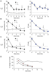
Bicuculline (10 µM) was appied at time 0. Arrows show the approximate time at which paroxysmal activity appeared. In all cases, once synchronized activity was established it became difficult to conduct further meaningful VmD measurements as the events increased in frequency and complexity. A. In L2 inhibition declined rapidly for the first 5–6 minutes before stabilizing at around 30–40% of control. Background excitation was largely unaffected. Despite this, the fall in estimated inhibition, the latter predominated throughout and the I∶E ratio never fell below 1. B. In L3 inhibition also fell rapidly over 5 minutes and continued to decline reaching a loss of around 85–90%. Simultaneously, excitation showed a gradual increase, although this did not reach significance. The combine effect was a sustained decrease in I∶E ration that reversed in favour of excitation around the time when paroxysmal activity appeared. C. Changes in background activity in L5 were similar to those in L3 except that the fall in inhibition was even more precipitous, and reached close to 100% in some cases. D. The time course of changes in I∶E ratio show that this occurred more rapidly in L5 than L3 but reached a similar end-point whereas that in L2 never reached the same level as that in either of the deeper layers.
Figure 7
Figure 7. Changes in variance of background conductances during bicuculline perfusion.
There were substantial differences in variances between and within layers, but scales have been adjusted to allow for comparison of the time course of changes. A. In L2 the variance (and thus synchronicity) of both inhibition and excitation fall rapidly over 2–3 minutes and thereafter remained reasonably stable at the lower level, although that of inhibition tended to increase again after the first fall. B. In L3 variance of inhibition fell steadily and remained stable whereas that of excitation showed little change. C. Similar to L3, inhibition variance fell throughout in L5, but an initial fall in variance of excitation was followed by a clear recovery to control levels. Again, the arrows indicate the approximate time at which paroxysmal activity appeared.
Figure 8
Figure 8. Additional effects of strychnine and glycine on paroxysmal activity in L2.
A. In the presence of bicuculline the spontaneous activity recorded in L2 consisted of single brief negative-gong events. Following addition of strychnine this was joined by a series of brief afterdischarges. A further, and more pronounced, exacerbation of the activity was seen when the non-competitive GABAAr antagonist, picrotoxin was added to the cocktail. B. A similar study in L2 of another slice shows that the same result was obtained when the order of application of strychnine and picrotoxin was reversed. C. In this slice we made simultaneous recordings at locations in L2, L3 and L5. Spontaneous events in bicuculline were briefer and less complex than those seen in the deeper layers (cf. Fig. 5). Addition of strychnine and then picrotoxin, as in the other studies increased the duration and complexity of events in L2, but left those in L3 and L5 largely unaltered.

Similar articles

Cited by

References

    1. Eichenbaum H (2001) The long and winding road to memory consolidation. Nat Neurosci 4: 1057–1058. - PubMed
    1. Squire LR, Stark CE, Clark RE (2004) The medial temporal lobe. Ann Rev Neurosci 27: 279–306. - PubMed
    1. Witter MP, Moser EI (2006) Spatial representation and the architecture of the entorhinal cortex. Trends Neurosci 29: 671–678. - PubMed
    1. Cunningham MO, Pervouchine DD, Racca C, Kopell NJ, Davies CH, et al. (2006) Neuronal metabolism governs cortical network response state. Proc Natl Acad Sci USA 103: 5597–5601. - PMC - PubMed
    1. Cunningham MO, Davies CH, Buhl EH, Kopell N, Whittington MA (2003) Gamma oscillations induced by kainate receptor activation in the entorhinal cortex in vitro. J Neurosci 23: 9761–9769. - PMC - PubMed

Publication types

LinkOut - more resources