

Comprehensive genetic analysis of early host body reactions to the bioactive and bio-inert porous scaffolds
- PMID: 24454803
- PMCID: PMC3891765
- DOI: 10.1371/journal.pone.0085132
Comprehensive genetic analysis of early host body reactions to the bioactive and bio-inert porous scaffolds
Abstract
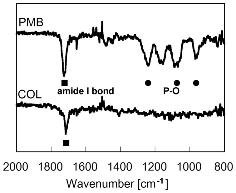
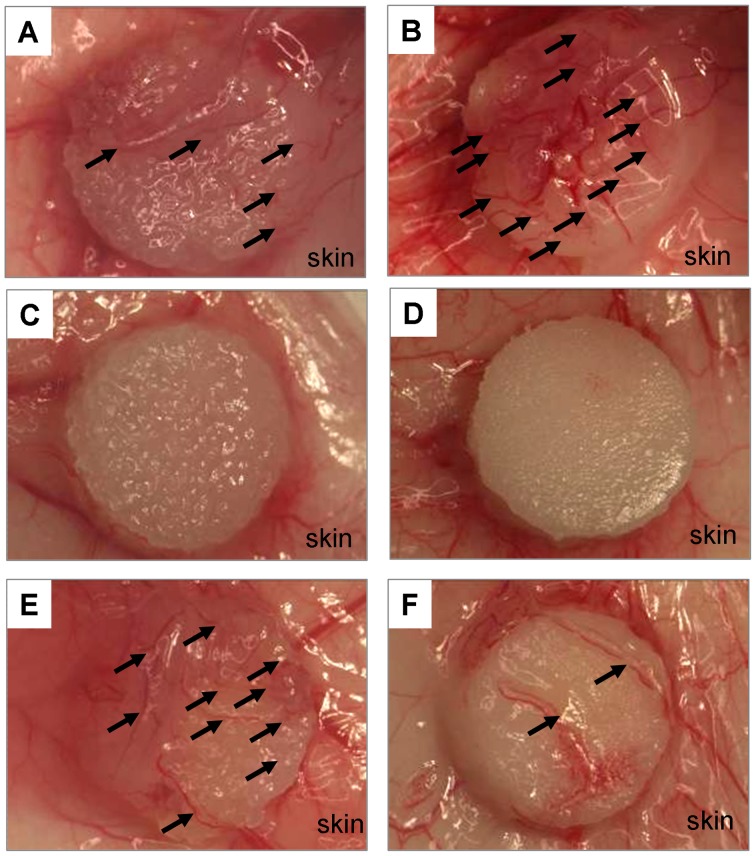
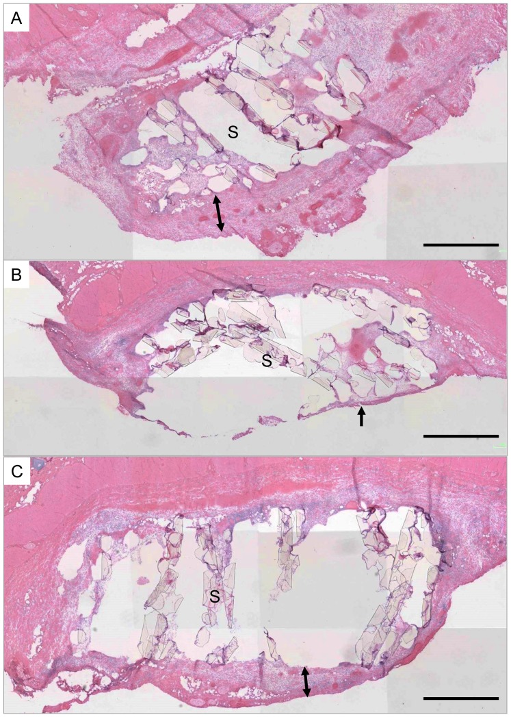
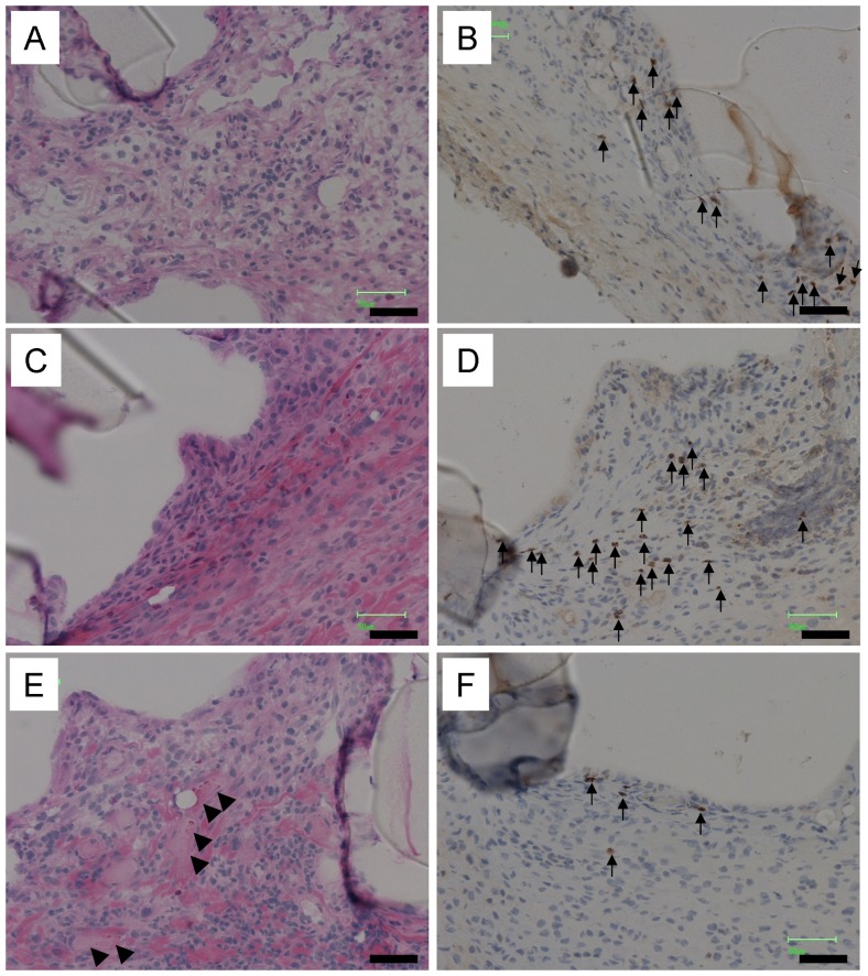
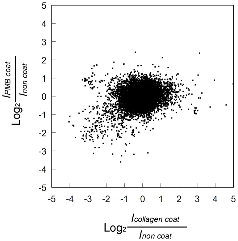
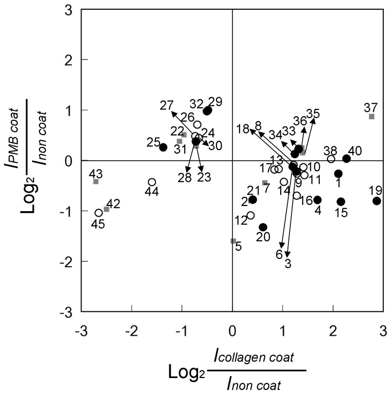
To design scaffolds for tissue regeneration, details of the host body reaction to the scaffolds must be studied. Host body reactions have been investigated mainly by immunohistological observations for a long time. Despite of recent dramatic development in genetic analysis technologies, genetically comprehensive changes in host body reactions are hardly studied. There is no information about host body reactions that can predict successful tissue regeneration in the future. In the present study, porous polyethylene scaffolds were coated with bioactive collagen or bio-inert poly(2-methacryloyloxyethyl phosphorylcholine-co-n-butyl methacrylate) (PMB) and were implanted subcutaneously and compared the host body reaction to those substrates by normalizing the result using control non-coat polyethylene scaffold. The comprehensive analyses of early host body reactions to the scaffolds were carried out using a DNA microarray assay. Within numerous genes which were expressed differently among these scaffolds, particular genes related to inflammation, wound healing, and angiogenesis were focused upon. Interleukin (IL)-1β and IL-10 are important cytokines in tissue responses to biomaterials because IL-1β promotes both inflammation and wound healing and IL-10 suppresses both of them. IL-1β was up-regulated in the collagen-coated scaffold. Collagen-specifically up-regulated genes contained both M1- and M2-macrophage-related genes. Marked vessel formation in the collagen-coated scaffold was occurred in accordance with the up-regulation of many angiogenesis-inducible factors. The DNA microarray assay provided global information regarding the host body reaction. Interestingly, several up-regulated genes were detected even on the very bio-inert PMB-coated surfaces and those genes include inflammation-suppressive and wound healing-suppressive IL-10, suggesting that not only active tissue response but also the inert response may relates to these genetic regulations.
Conflict of interest statement
Figures
Similar articles
-
Gene chip/PCR-array analysis of tissue response to 2-methacryloyloxyethyl phosphorylcholine (MPC) polymer surfaces in a mouse subcutaneous transplantation system.J Biomater Sci Polym Ed. 2014;25(14-15):1658-72. doi: 10.1080/09205063.2014.939917. Epub 2014 Jul 30. J Biomater Sci Polym Ed. 2014. PMID: 25075735
-
The effect of cell debris within biologic scaffolds upon the macrophage response.J Biomed Mater Res A. 2017 Aug;105(8):2109-2118. doi: 10.1002/jbm.a.36055. Epub 2017 Apr 12. J Biomed Mater Res A. 2017. PMID: 28263432
-
Evaluation of in vitro macrophage response and in vivo host response to growth factors incorporated chitosan nanoparticle impregnated collagen-chitosan scaffold.J Biomed Nanotechnol. 2014 Mar;10(3):508-18. doi: 10.1166/jbn.2014.1714. J Biomed Nanotechnol. 2014. PMID: 24730246
-
Copolymers of 2-methacryloyloxyethyl phosphorylcholine (MPC) as biomaterials.Biomed Mater Eng. 2004;14(4):345-54. Biomed Mater Eng. 2004. PMID: 15472384 Review.
-
Chitosan and fish collagen as biomaterials for regenerative medicine.Adv Food Nutr Res. 2012;65:107-20. doi: 10.1016/B978-0-12-416003-3.00006-8. Adv Food Nutr Res. 2012. PMID: 22361182 Review.
Cited by
-
Cell population kinetics of collagen scaffolds in ex vivo oral wound repair.PLoS One. 2014 Nov 14;9(11):e112680. doi: 10.1371/journal.pone.0112680. eCollection 2014. PLoS One. 2014. PMID: 25397671 Free PMC article.
-
Wound Healing: Biologics, Skin Substitutes, Biomembranes and Scaffolds.Healthcare (Basel). 2014 Sep 10;2(3):356-400. doi: 10.3390/healthcare2030356. Healthcare (Basel). 2014. PMID: 27429283 Free PMC article. Review.
-
Immunomodulation of Skin Repair: Cell-Based Therapeutic Strategies for Skin Replacement (A Comprehensive Review).Biomedicines. 2022 Jan 6;10(1):118. doi: 10.3390/biomedicines10010118. Biomedicines. 2022. PMID: 35052797 Free PMC article. Review.
-
Delivery strategies to control inflammatory response: Modulating M1-M2 polarization in tissue engineering applications.J Control Release. 2016 Oct 28;240:349-363. doi: 10.1016/j.jconrel.2016.01.026. Epub 2016 Jan 14. J Control Release. 2016. PMID: 26778695 Free PMC article. Review.
-
Bioengineered Corneas Grafted as Alternatives to Human Donor Corneas in Three High-Risk Patients.Clin Transl Sci. 2015 Oct;8(5):558-62. doi: 10.1111/cts.12293. Epub 2015 May 21. Clin Transl Sci. 2015. PMID: 25996570 Free PMC article.
References
-
- Nel AE, Mädler L, Velegol D, Xia T, Hoek EMV, et al. (2009) Understanding biophysicochemical interactions at the nano-bio interface. Nat Mater 8: 543–557. - PubMed
-
- Matheson LA, Naksym GN, Santerre JP, Labow RS (2006) Cyclic biaxial strain affects U937 macrophage-like morphology and enzymatic activities. J Biomed Mater Res A 76: 52–62. - PubMed
-
- Yeo Y, Bellas E, Highley CB, Langer R, Kohane DS (2007) Peritoneal adhesion prevention with an in situ cross-linkable hyaluronan gel containing tissue-type plasminogen activator in a rabbit repeated-injury model. Biomaterials 28: 3704–3713. - PubMed
Publication types
MeSH terms
Substances
LinkOut - more resources
Full Text Sources
Other Literature Sources
Molecular Biology Databases
