

Durable remission of renal cell carcinoma in conjuncture with graft versus host disease following allogeneic stem cell transplantation and donor lymphocyte infusion: rule or exception?
- PMID: 24454818
- PMCID: PMC3893183
- DOI: 10.1371/journal.pone.0085198
Durable remission of renal cell carcinoma in conjuncture with graft versus host disease following allogeneic stem cell transplantation and donor lymphocyte infusion: rule or exception?
Abstract
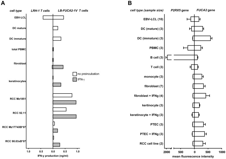
Allogeneic stem cell transplantation (alloSCT) followed by donor lymphocyte infusion (DLI) can be applied as immunotherapeutic intervention to treat malignant diseases. Here, we describe a patient with progressive metastatic clear cell renal cell carcinoma (RCC) who was treated with T cell depleted non-myeloablative alloSCT and DLI resulting in disease regression accompanied by extensive graft versus host disease (GVHD). We characterized the specificity of this immune response, and detected a dominant T cell population recognizing a novel minor histocompatibility antigen (MiHA) designated LB-FUCA2-1V. T cells specific for LB-FUCA2-1V were shown to recognize RCC cell lines, supporting a dominant role in the graft versus tumor (GVT) reaction. However, coinciding with the gradual disappearance of chronic GVHD, the anti-tumor effect declined and 3 years after alloSCT the metastases became progressive again. To re-initiate the GVT reaction, escalating doses of DLI were given, but no immune response could be induced and the patient died of progressive disease 8.5 years after alloSCT. Gene expression studies illustrated that only a minimal number of genes shared expression between RCC and professional antigen presenting cells but were not expressed by non-malignant healthy tissues, indicating that in patients suffering from RCC, GVT reactivity after alloSCT may be unavoidably linked to GVHD.
Conflict of interest statement
Figures


Similar articles
-
CD4 Donor Lymphocyte Infusion Can Cause Conversion of Chimerism Without GVHD by Inducing Immune Responses Targeting Minor Histocompatibility Antigens in HLA Class II.Front Immunol. 2018 Dec 18;9:3016. doi: 10.3389/fimmu.2018.03016. eCollection 2018. Front Immunol. 2018. PMID: 30619360 Free PMC article.
-
Alloreactivity as therapeutic principle in the treatment of hematologic malignancies. Studies of clinical and immunologic aspects of allogeneic hematopoietic cell transplantation with nonmyeloablative conditioning.Dan Med Bull. 2007 May;54(2):112-39. Dan Med Bull. 2007. PMID: 17521527 Review.
-
A polymorphism in the splice donor site of ZNF419 results in the novel renal cell carcinoma-associated minor histocompatibility antigen ZAPHIR.PLoS One. 2011;6(6):e21699. doi: 10.1371/journal.pone.0021699. Epub 2011 Jun 28. PLoS One. 2011. PMID: 21738768 Free PMC article.
-
Tissue Damage Caused by Myeloablative, but Not Non-Myeloablative, Conditioning before Allogeneic Stem Cell Transplantation Results in Dermal Macrophage Recruitment without Active T-Cell Interaction.Front Immunol. 2018 Feb 27;9:331. doi: 10.3389/fimmu.2018.00331. eCollection 2018. Front Immunol. 2018. PMID: 29535719 Free PMC article. Clinical Trial.
-
Donor lymphocyte infusion: the use of alloreactive and tumor-reactive lymphocytes for immunotherapy of malignant and nonmalignant diseases in conjunction with allogeneic stem cell transplantation.J Hematother Stem Cell Res. 2002 Apr;11(2):265-76. doi: 10.1089/152581602753658457. J Hematother Stem Cell Res. 2002. PMID: 11983098 Review.
Cited by
-
Identification of potential pathogenic biomarkers in clear cell renal cell carcinoma.Oncol Lett. 2018 Jun;15(6):8491-8499. doi: 10.3892/ol.2018.8398. Epub 2018 Mar 30. Oncol Lett. 2018. PMID: 29805586 Free PMC article.
-
Autosomal Minor Histocompatibility Antigens: How Genetic Variants Create Diversity in Immune Targets.Front Immunol. 2016 Mar 15;7:100. doi: 10.3389/fimmu.2016.00100. eCollection 2016. Front Immunol. 2016. PMID: 27014279 Free PMC article. Review.
-
An unexplored angle: T cell antigen discoveries reveal a marginal contribution of proteasome splicing to the immunogenic MHC class I antigen pool.Proc Natl Acad Sci U S A. 2022 Jul 19;119(29):e2119736119. doi: 10.1073/pnas.2119736119. Epub 2022 Jul 8. Proc Natl Acad Sci U S A. 2022. PMID: 35858315 Free PMC article.
-
The Connection Between Minor H Antigens and Neoantigens and the Missing Link in Their Prediction.Front Immunol. 2020 Jun 24;11:1162. doi: 10.3389/fimmu.2020.01162. eCollection 2020. Front Immunol. 2020. PMID: 32670277 Free PMC article. Review.
References
-
- Appelbaum FR (2003) The current status of hematopoietic cell transplantation. Annu Rev Med 54: 491–512. - PubMed
-
- von dem Borne PA, Beaumont F, Starrenburg CW, Oudshoorn M, Hale G, et al. (2006) Outcomes after myeloablative unrelated donor stem cell transplantation using both in vitro and in vivo T-cell depletion with alemtuzumab. Haematologica 91: 1559–1562. - PubMed
-
- Childs R, Chernoff A, Contentin N, Bahceci E, Schrump D, et al. (2000) Regression of metastatic renal-cell carcinoma after nonmyeloablative allogeneic peripheral-blood stem-cell transplantation. N Engl J Med 343: 750–758. - PubMed
Publication types
MeSH terms
LinkOut - more resources
Full Text Sources
Other Literature Sources
Medical
Miscellaneous
