. 2014 Jan 15;9(1):e85511.
doi: 10.1371/journal.pone.0085511. eCollection 2014.

Evidence of spondyloarthropathy in the spine of a phytosaur (Reptilia: Archosauriformes) from the Late Triassic of Halberstadt, Germany

Affiliations

Evidence of spondyloarthropathy in the spine of a phytosaur (Reptilia: Archosauriformes) from the Late Triassic of Halberstadt, Germany

Florian Witzmann et al. PLoS One. .

Abstract

Pathologies in the skeleton of phytosaurs, extinct archosauriform reptiles restricted to the Late Triassic, have only been rarely described. The only known postcranial pathologies of a phytosaur are two pairs of fused vertebrae of "Angistorhinopsis ruetimeyeri" from Halberstadt, Germany, as initially described by the paleontologist Friedrich von Huene. These pathologic vertebrae are redescribed in more detail in this study in the light of modern paleopathologic methods. Four different pathologic observations can be made in the vertebral column of this individual: 1) fusion of two thoracic vertebral bodies by new bone formation within the synovial membrane and articular capsule of the intervertebral joint; 2) fusion and conspicuous antero-posterior shortening of last presacral and first sacral vertebral bodies; 3) destruction and erosion of the anterior articular surface of the last presacral vertebra; and 4) a smooth depression on the ventral surface of the fused last presacral and first sacral vertebral bodies. Observations 1-3 can most plausibly and parsimoniously be attributed to one disease: spondyloarthropathy, an aseptic inflammatory process in which affected vertebrae show typical types of reactive new bone formation and erosion of subchondral bone. The kind of vertebral shortening present in the fused lumbosacral vertebrae suggests that the phytosaur acquired this disease in its early life. Observation 4, the smooth ventral depression in the fused lumbosacral vertebrae, is most probably not connected to the spondyloarthropathy, and can be regarded as a separate abnormality. It remains of uncertain origin, but may be the result of pressure, perhaps caused by a benign mass such as an aneurysm or cyst of unknown type. Reports of spondyloarthropathy in Paleozoic and Mesozoic reptiles are still exceptional, and our report of spondyloarthropathy in fossil material from Halberstadt is the first unequivocal occurrence of this disease in a Triassic tetrapod and in a phytosaur.


Conflict of interest statement

Competing Interests: The authors have declared that no competing interests exist.

Figures

Figure 1
Figure 1. Reconstruction of a phytosaur skeleton (Rutiodon), redrawn from .
Phytosaurs superficially resemble crocodiles in their habitus and probably had a similar amphibious lifestyle and ecology.
Figure 2
Figure 2. Morphology of “normal” phytosaur vertebrae (Nicrosaurus kapffi).
A, B. SMNS 5999, left lateral view of presacral vertebra. C. SMNS 4384, posterior presacral “lumbar” vertebra in anterior view. Abbreviations: aas, anterior articular surface; cdf, centrodiapophyseal fossa; fov, fovea; ipof, infrapostzygapophyseal fossa; nc, neural canal; nsp, neural spine; par, parapophysis; pcdl, posterior centrodiapophyseal lamina; poz, postzygapophysis; ppdl, paradiapophyseal lamina; prz, prezygapophysis; trp, transverse process.
Figure 3
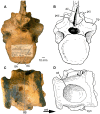
Figure 3. Phytosauria indet. from the Late Triassic of Halberstadt. Fused vertebrae MB.R.1972 derived from the thoracic region.
A, B. Right lateral view. C, D. Ventral view. E, F. Dorsal view. The dashed lines in (A) show the location of sections illustrated in Figure 7C, D. Abbreviations: acdl, anterior centrodiapophyseal lamina; ant, anterior; bu, bulge; but, buttress; cdf, centrodiapophyseal fossa; dia, diapophysis; ep, epipophys; nf, nutrient foramina; nsp, neural spine; par, parapophysis; pcdl, posterior centrodiapophyseal lamina; pocdf, postzygapophyseal centrodiapophyseal fossa; podl, postzygodiapophyseal lamina; poz, postzygapophysis; ppdl, paradiapophyseal lamina; prcdf, prezygapophyseal centrodiapophyseal fossa; prdl, prezygodiapophyseal lamina; prz, prezygapophysis; sprf, spinoprezygapophyseal fossa; sprl, spinoprezygapophyseal lamina; trp, transverse process.
Figure 4
Figure 4. Phytosauria indet. from the Late Triassic of Halberstadt. Fused vertebrae MB.R.1972 derived from the thoracic region.
A, B. Anterior view. C, D. Posterior view. The dashed lines in (C) show the location of sections illustrated in Figure 7A, B. Abbreviations: cdf, centrodiapophyseal fossa; cpol, centropostzygapophyseal lamina; cprl, centroprezygapophyseal lamina; dia, diapophysis; nc, neural canal; nsp, neural spine; par, parapophysis; pocdf, postzygapophyseal centrodiapophyseal fossa; poz, postzygapophysis; prcdf, prezygapophyseal centrodiapophyseal fossa; prz, prezygapophyses; spof, spinopostzygapophyseal fossa; spol, spinopostzygapophyseal laminae; sprf, spinoprezygapophyseal fossa; trp, transverse process.
Figure 5
Figure 5. Phytosauria indet. from the Late Triassic of Halberstadt. Fused vertebrae MB.R.1973 derived from the lumbosacral region.
A, B. Right lateral view. C. Anterior endplate showing severe destruction of its surface. D. Close up of the ventral part of the anterior endplate. Abbreviations: acdl, anterior centrodiapophyseal lamina; ant, anterior; cdf, centrodiapophyseal fossa; dia, diapophysis; nc, neural canal; nsp, neural spine; pcdl, posterior centrodiapophyseal lamina; poz, postzygapophysis; prz, prezygapophysis; syn, synapophysis; trp, transverse process.
Figure 6
Figure 6. Phytosauria indet. from the Late Triassic of Halberstadt. Fused vertebrae MB.R.1973 derived from the lumbosacral region.
A, B. Posterior view. C, D. Ventral view, showing ovate depression on the ventral surface of the synostosed vertebral bodies. The dashed lines in (A) show the location of sections illustrated in Figure 8A, C; the dashed line in (C) shows the location of section illustrated in Figure 8 B. Abbreviations: ant, anterior; dil, depression for interspinous ligament; nc, neural canal; od, ovate depression; poz, postzygapophysis; prz, prezygapophysis; syn, synapophysis; trp, transverse process.
Figure 7
Figure 7. Phytosauria indet. from the Late Triassic of Halberstadt. CT scans of the fused vertebrae MB.R.1972.
A. Sagittal section along the midline of the vertebrae. B. Sagittal section through the left lateral part of the vertebrae. C. Transverse section through the anterior vertebra. D. Transverse section through the intervertebral space, showing a ring-like structure. For the location of sections, please see Figures 3A and 4C. Abbreviations: ivs, intervertebral space; nc, neural canal; nsp, neural spine; trp, transverse process.
Figure 8
Figure 8. Phytosauria indet. from the Late Triassic of Halberstadt. CT scans of the fused vertebrae MB.R.1973.
A. Sagittal section along the midline of the vertebrae. B. Transverse section through the intervertebral space, showing a simple cavity or lacuna. C. Sagittal section through the right lateral part of the vertebrae, showing their complete fusion. For the location of sections, please see Figure 6A, C. Abbreviations: aas, anterior articular surface; ivs, intervertebral space; nc, neural canal; nsp, neural spine; od, ovate depression; prz, prezygapophyses.
Figure 9
Figure 9. Schematic drawing of the synovial articulation between procoelous thoracic vertebral bodies of an extant crocodylian.
Redrawn after ; articular cartilage covering the vertebral articular surfaces is added, based on . Abbreviations: aas, anterior articular surface (vertebral fossa); acap, articular capsule; acart, articular cartilage; anul, ring of fibrous cartilage; fov, fovea; pas, posterior articular surface (vertebral condyle); peo, periost; sept, intercorporal septum; syns, synovial space.

Similar articles

Cited by

References

    1. Hungerbühler A (2002) The Late Triassic phytosaur Mystriosuchus westphali, with a revision of the genus. Palaeontology 45: 377–418.
    1. Stocker MR (2012) A new phytosaur (Archosauriformes, Phytosauria) from the Lot’s Wife beds (Sonsela Member) within the Chinle Formation (Upper Triassic) of Petrified Forest National Park, Arizona. J Vert Paleontol 32: 573–586.
    1. McGregor JH (1906) The Phytosauria, with especial reference to Mystriosuchus and Rhytidodon . Mem Am Mus Nat Hist 9: 27–100.
    1. Camp CL (1930) A study of the phytosaurs with description of new material from western North America. Mem Univ Calif 10: 1–174.
    1. Case EC (1932) A perfectly preserved segment of the armor of a phytosaur, with associated vertebrae. Contrib Mus Paleontol Univ Mich 4 (2): 57–80.

Publication types

LinkOut - more resources