. 2014 Jan 16;9(1):e85765.
doi: 10.1371/journal.pone.0085765. eCollection 2014.

Antimicrobial cathelicidin peptide LL-37 inhibits the LPS/ATP-induced pyroptosis of macrophages by dual mechanism

Affiliations

Antimicrobial cathelicidin peptide LL-37 inhibits the LPS/ATP-induced pyroptosis of macrophages by dual mechanism

Zhongshuang Hu et al. PLoS One. .

Abstract

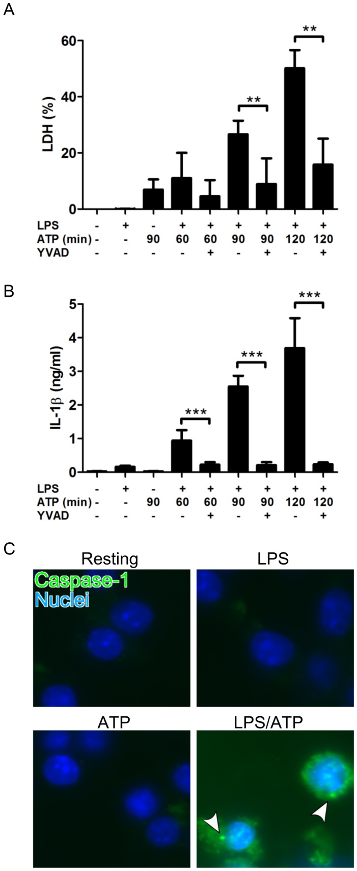
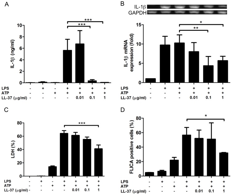
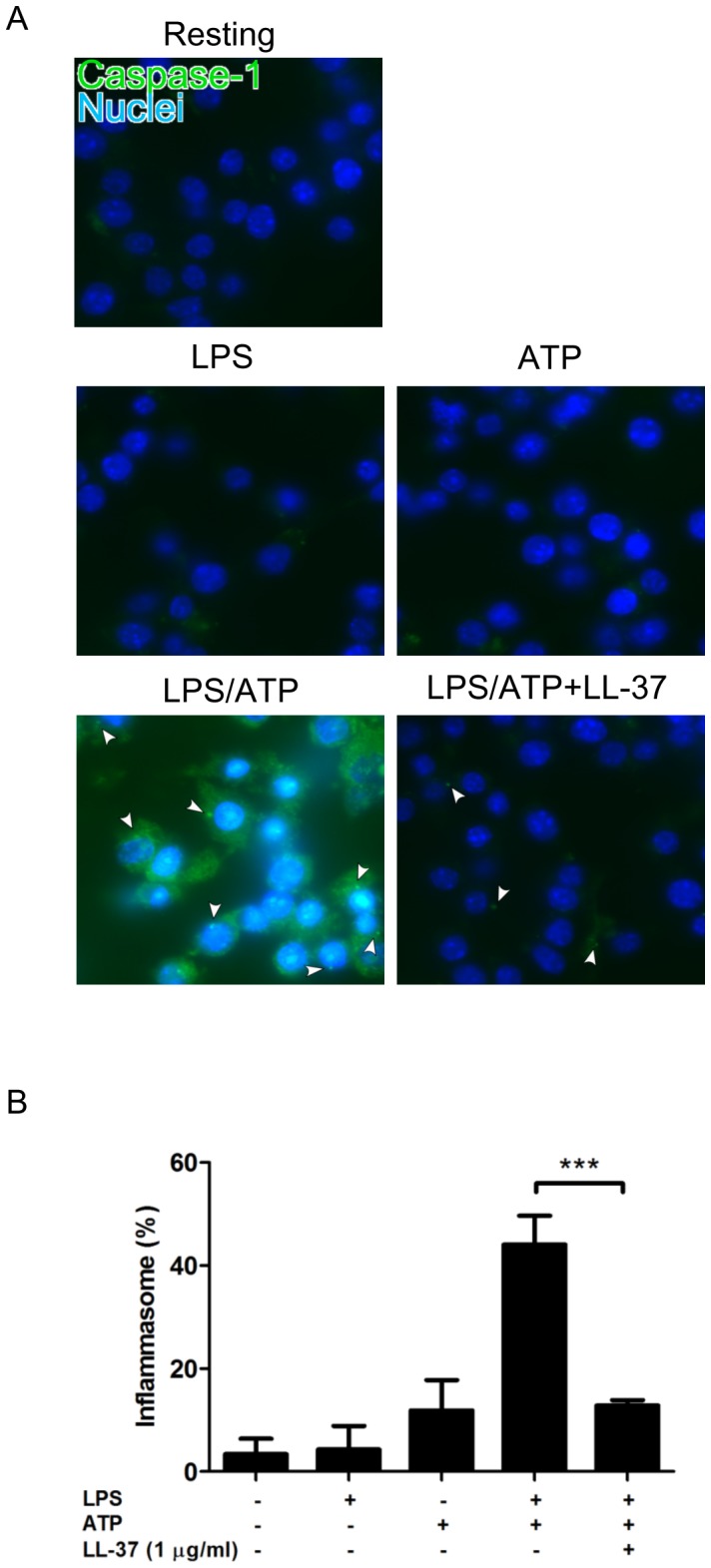
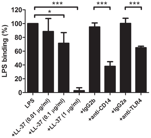
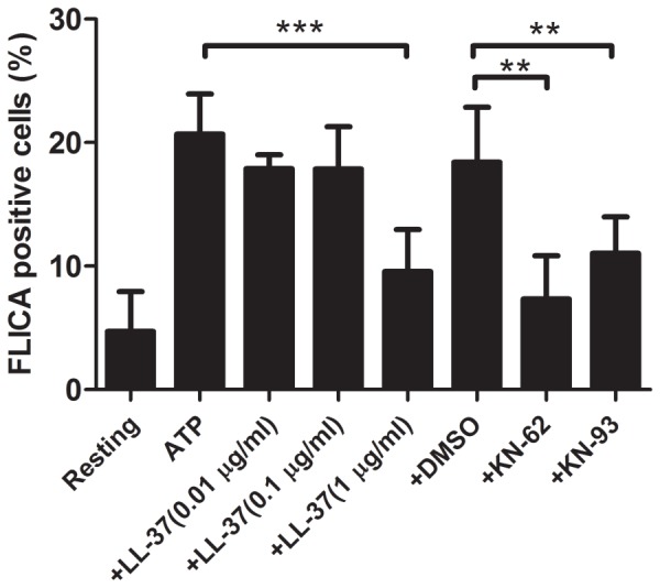
Pyroptosis is a caspase-1 dependent cell death, associated with proinflammatory cytokine production, and is considered to play a crucial role in sepsis. Pyroptosis is induced by the two distinct stimuli, microbial PAMPs (pathogen associated molecular patterns) and endogenous DAMPs (damage associated molecular patterns). Importantly, cathelicidin-related AMPs (antimicrobial peptides) have a role in innate immune defense. Notably, human cathelicidin LL-37 exhibits the protective effect on the septic animal models. Thus, in this study, to elucidate the mechanism for the protective action of LL-37 on sepsis, we utilized LPS (lipopolysaccharide) and ATP (adenosine triphosphate) as a PAMP and a DAMP, respectively, and examined the effect of LL-37 on the LPS/ATP-induced pyroptosis of macrophage-like J774 cells. The data indicated that the stimulation of J774 cells with LPS and ATP induces the features of pyroptosis, including the expression of IL-1β mRNA and protein, activation of caspase-1, inflammasome formation and cell death. Moreover, LL-37 inhibits the LPS/ATP-induced IL-1β expression, caspase-1 activation, inflammasome formation, as well as cell death. Notably, LL-37 suppressed the LPS binding to target cells and ATP-induced/P2X7-mediated caspase-1 activation. Together these observations suggest that LL-37 potently inhibits the LPS/ATP-induced pyroptosis by both neutralizing the action of LPS and inhibiting the response of P2X7 to ATP. Thus, the present finding may provide a novel insight into the modulation of sepsis utilizing LL-37 with a dual action on the LPS binding and P2X7 activation.


Conflict of interest statement

Competing Interests: The authors have declared that no competing interests exist.

Figures

Figure 1
Figure 1. Effect of LPS and ATP treatment on the pyroptosis of J774 cells.
Macrophage-like J774 cells were primed with 10 ng/ml LPS for 4 h, and then treated with 3 mM ATP for the indicated time periods in the absence or presence of 20 µM Ac-YVAD-CHO, a caspase-1 specific inhibitor. Cells were also incubated with LPS or ATP alone, or without LPS and ATP (Resting). Thereafter, the supernatants were recovered for the assays of IL-1β (A) and LDH (B). IL-1β 1evels were determined using a commercially available mouse IL-1β ELISA kit. LDH activities in the supernatants and 1% Triton X-100-lysed cells (as a total activity of 100%) were determined using a commercially available LDH assay kit. Data shows the mean ± SD of 3 separate experiments. Values are compared between the absence and presence of Ac-YVAD-CHO. **P<0.01, ***P< 0.001. (C) J774 cells were primed with 10 ng/ml LPS for 4 h and then treated with 3 mM ATP for 90 min. Thereafter, the cells were stained with FAM-YVAD-fmk (a fluorescent labeled caspase-1 inhibitor for inflammasome staining, green) and Hoechst 33342 (for nuclear staining, blue) and photographed with a fluorescence microscope system. Arrowheads indicate the inflammasomes containing the activated caspase-1. Images of cells are representative of 3 separate experiments.
Figure 2
Figure 2. Effect of LL-37 on the LPS/ATP-induced pyroptosis of J774 cells.
J774 cells were primed with 10/ml LPS for 4 h, and then treated with 3 mM ATP for 90 min in the absence or presence of LL-37 (0.01, 0.1 or 1 µg/ml). Cells were also incubated with LPS or ATP alone, or without LPS and ATP. Thereafter, the supernatants were recovered for the assays of IL-1β (A) and LDH (C), and the cells were used for the assays of IL-1β mRNA expression (B) and caspase-1 activation (D). IL-1β mRNA expression were determined by RT-PCR and expressed as fold increase relative to resting cells incubated without LPS, ATP and LL-37; the caspase-1 activation was assayed by flow cytometry using FAM-YVAD-fmk (a fluorescent labeled inhibitor of caspase-1, FLICA) that irreversibly binds with activated caspase-1, and expressed as the percentage of FLICA positive cells. Data shows the mean ± SD of 3-5 separate experiments. Values are compared between the absence and presence of LL-37 among LPS/ATP-treated cells. *P<0.05, **P<0.01, ***P<0.001. Images of RT-PCR are representative of 3-5 separate experiments.
Figure 3
Figure 3. Effect of LL-37 on the LPS/ATP-induced inflammasome formation in J774 cells.
J774 cells were primed with 10/ml LPS for 4 h, and then treated with 3 mM ATP for 90 min in the absence or presence of LL-37 (1 µg/ml). Cells were also incubated with LPS or ATP alone, or without LPS and ATP (Resting). Thereafter, the cells were stained with FAM-YVAD-fmk (a fluorescent labeled caspase-1 inhibitor) and Hoechst 33342 (A). Arrowheads indicate the inflammasomes containing the activated caspase-1. Furthermore, the percentage of inflammasome-containing cells with activated caspase-1 was determined by counting at least 200 Hoechst positive cells (B). Data shows the mean ± SD of 3-5 separate experiments. Values are compared in the LPS/ATP-treated cells between the absence and presence of LL-37. ***P<0.001. Images cells are representative of 3–5 separate experiments.
Figure 4
Figure 4. Effect of LL-37 on the LPS binding to J774 cells.
J774 cells were suspended in DMEM containing 10% FBS, and incubated with 1 µg/ml FITC-LPS at 37°C for 15 min in the absence or presence of LL-37 (0.01, 0.1 or 1 µg/ml), anti-mouse CD14 monoclonal antibody (4C1, 10 µg/ml), anti-mouse TLR4 monoclonal antibody (MTS510, 40 µg/ml) or isotype control IgG (IgG2b and IgG2a). The binding of LPS was analyzed by flow cytometry, and the median fluorescence intensity was determined. The LPS binding was expressed as the percentage of that with FITC-LPS alone. Data shows the mean ± SD of 3 separate experiments. Values are compared between the absence and presence of LL-37, anti-CD14 monoclonal antibody or anti-TLR4 monoclonal antibody. **P<0.01, ***P<0.001.
Figure 5
Figure 5. Effect of LL-37 on the ATP-induced caspase-1 activation in J774 cells.
J774 cells were treated with 3-37 (0.01, 0.1 or 1 µg/ml), P2X7 antagonists (1 µM KN-62 and 1 µM KN-93) or dimethylsulfoxide (DMSO, a solvent for KN-62 and KN-93, 0.1%). Cells were also incubated without ATP, LL-37 and P2X7 antagonists (Resting). Thereafter, the caspase-1 activation was assayed by flow cytometry using FAM-YVAD-fmk (FLICA), and expressed as the percentage of FLICA positive cells. Data shows the mean ± SD of 3 separate experiments. Values are compared between the absence and presence of LL-37 or P2X7 antagonists among ATP-treated cells. **P<0.01, ***P<0.001.

Similar articles

Cited by

References

    1. Bone RC, Balk RA, Cerra FB, Dellinger RP, Fein AM, et al. (1992) Definitions for sepsis and organ failure and guidelines for the use of innovative therapies in sepsis. The ACCP/SCCM Consensus Conference Committee. American College of Chest Physicians/Society of Critical Care Medicine. Chest 101: 1644–1655. - PubMed
    1. Angus DC (2011) The search for effective therapy for sepsis: back to the drawing board? Jama 306: 2614–2615. - PubMed
    1. O′Brien JM Jr, Ali NA, Aberegg SK, Abraham E (2007) Sepsis. Am J Med 120: 1012–1022. - PubMed
    1. Levy MM, Dellinger RP, Townsend SR, Linde-Zwirble WT, Marshall JC, et al. (2011) The Surviving Sepsis Campaign: results of an international guideline-based performance improvement program targeting severe sepsis. Crit Care Med 38: 367–374. - PubMed
    1. Pinsky MR (2004) Dysregulation of the immune response in severe sepsis. Am J Med Sci 328: 220–229. - PubMed

Publication types

LinkOut - more resources