. 2014 Jan 15;9(1):e86146.
doi: 10.1371/journal.pone.0086146. eCollection 2014.

Production of pigs expressing a transgene under the control of a tetracycline-inducible system

Affiliations

Production of pigs expressing a transgene under the control of a tetracycline-inducible system

Yong-Xun Jin et al. PLoS One. .

Abstract

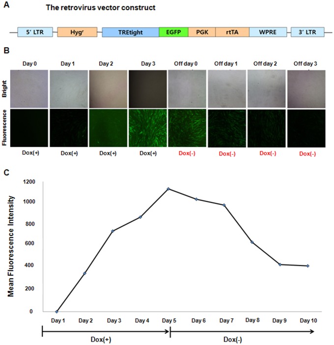
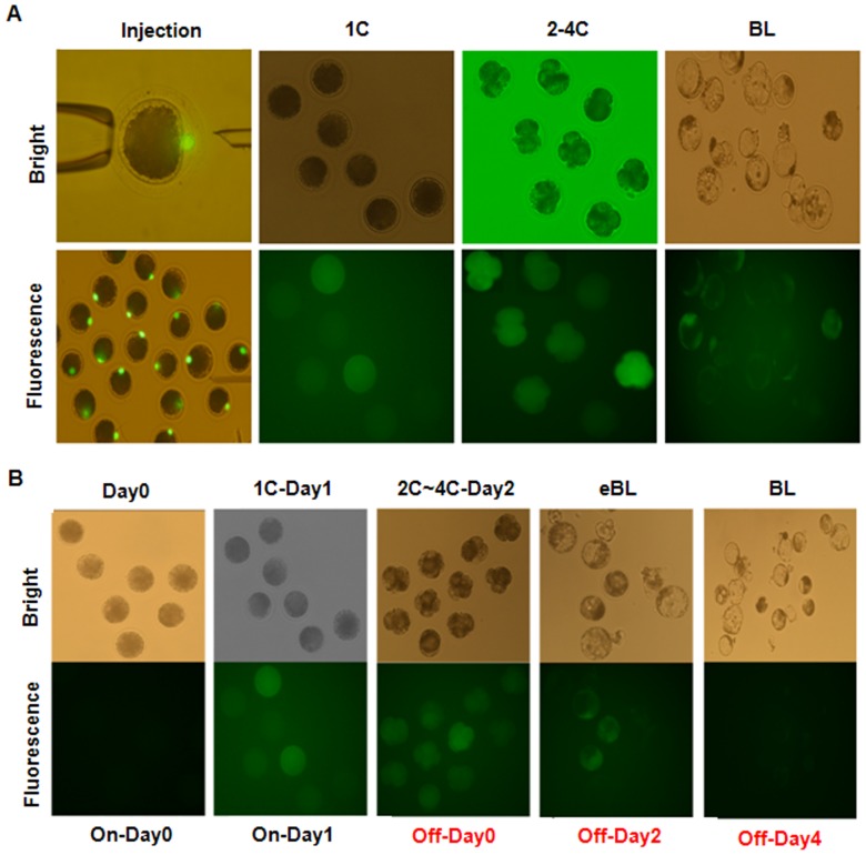
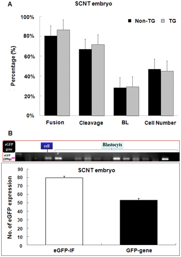
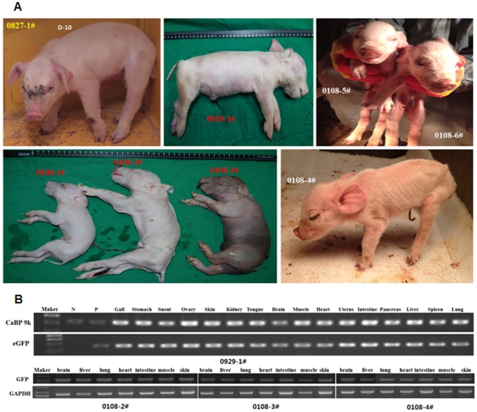
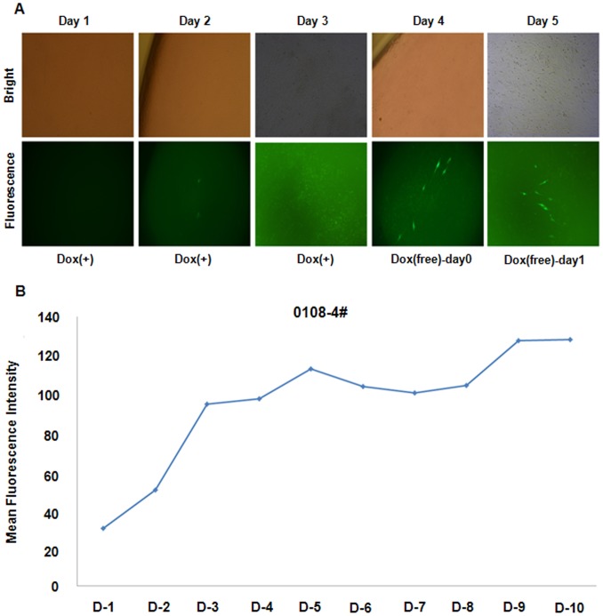
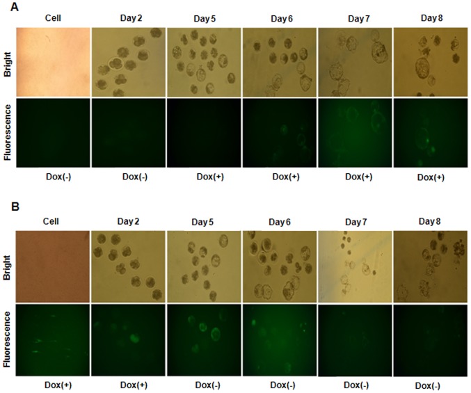
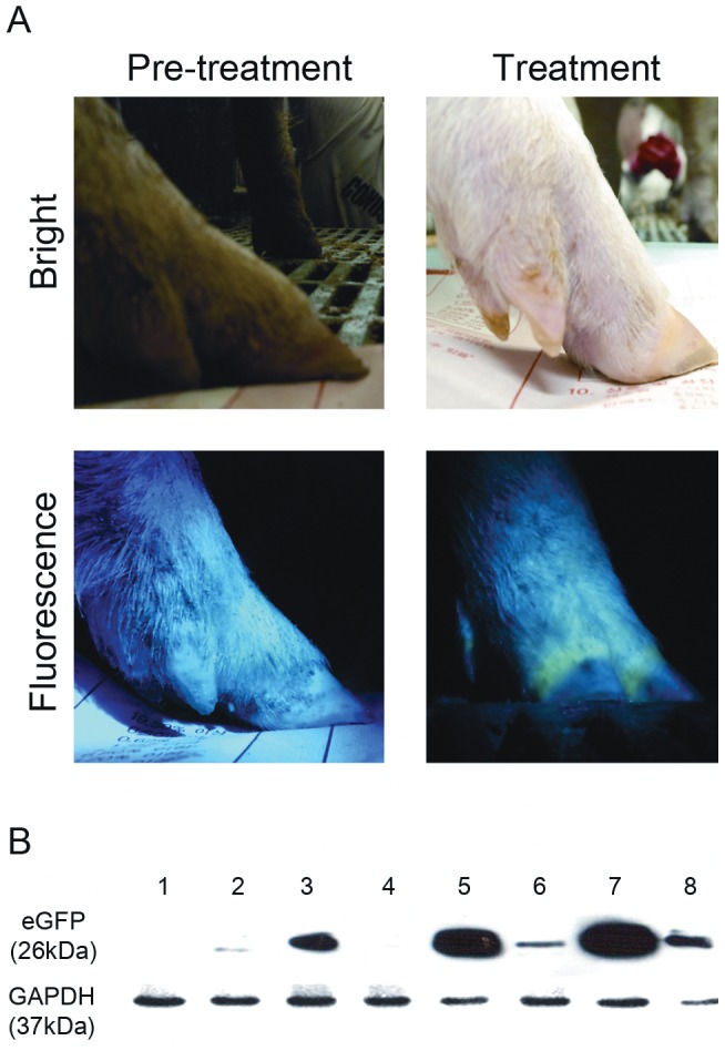
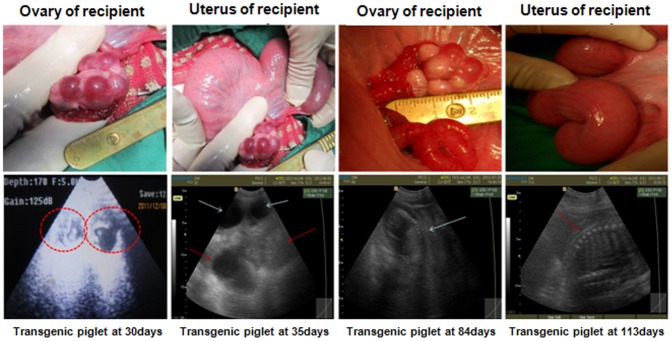
Pigs are anatomically and physiologically closer to humans than other laboratory animals. Transgenic (TG) pigs are widely used as models of human diseases. The aim of this study was to produce pigs expressing a tetracycline (Tet)-inducible transgene. The Tet-on system was first tested in infected donor cells. Porcine fetal fibroblasts were infected with a universal doxycycline-inducible vector containing the target gene enhanced green fluorescent protein (eGFP). At 1 day after treatment with 1 µg/ml doxycycline, the fluorescence intensity of these cells was increased. Somatic cell nuclear transfer (SCNT) was then performed using these donor cells. The Tet-on system was then tested in the generated porcine SCNT-TG embryos. Of 4,951 porcine SCNT-TG embryos generated, 850 were cultured in the presence of 1 µg/ml doxycycline in vitro. All of these embryos expressed eGFP and 15 embryos developed to blastocyst stage. The remaining 4,101 embryos were transferred to thirty three surrogate pigs from which thirty eight cloned TG piglets were obtained. PCR analysis showed that the transgene was inserted into the genome of each of these piglets. Two TG fibroblast cell lines were established from these TG piglets, and these cells were used as donor cells for re-cloning. The re-cloned SCNT embryos expressed the eGFP transgene under the control of doxycycline. These data show that the expression of transgenes in cloned TG pigs can be regulated by the Tet-on/off systems.


Conflict of interest statement

Competing Interests: The authors have declared that no competing interests exist.

Figures

Figure 1
Figure 1. Induction of eGFP expression by doxycycline in Tet-on-eGFP pFF cells.
(A) Schematic diagram of the retrovirus construct. LTR, long terminal repeat; HygR, hygromycin B resistance gene; TRE-tight, modified version of the tetracycline-response element; mCMV, minimal cytomegalovirus promoter; eGFP, enhanced green fluorescent protein; PGK, phosphoglycerate kinase promoter; rtTA2s-M2, reverse tetracycline transactivator; and WPRE, woodchuck hepatitis virus post-transcriptional regulatory element sequence. (B) Tet-on-eGFP pFF were cultured in the presence of doxycycline (Dox, 1 µg/ml) for up to 3 days and then in the absence of doxycycline for up to 3 days. The embryos were examined by bright field and fluorescence microscopy. (C) Tet-on-eGFP pFF were cultured in the presence of doxycycline (1 µg/ml) for up to 5 days and then in the absence of doxycycline for a further 1–5 days. The mean fluorescence intensity of the cells was assessed by FACS.
Figure 2
Figure 2. SCNT-TG embryos express eGFP when cultured in the presence of doxycycline.
(A) SCNT was performed using eGFP-positive pFF. The resulting embryos were cultured in the presence of doxycycline for 1 to 7 days and were examined by bright field and fluorescence microscopy. eGFP expression was detected in embryos at the 1-cell, 2–4-cell, and blastocyst stages. (B) SCNT-TG embryos were cultured in the presence of doxycycline for 3 days and then in the absence of doxycycline for a further 4 days. The embryos were examined by bright field and fluorescence microscopy. eGFP fluorescence appeared when embryos were cultured in the presence of doxycycline and gradually disappeared when embryos were cultured in the absence of doxycycline.
Figure 3
Figure 3. The development of SCNT embryos is similar regardless of whether they receive eGFP-positive pFF or control pFF.
(A) The fusion rate, cleavage rate, percentage of embryos that developed to blastocyst stage, and doxycycline were compared between SCNT embryos that received eGFP-positive pFF and those that received control pFF (non-TG). (B) PCR analyses confirmed that the eGFP gene was integrated into the genome of each blastocyst. Genomic DNA extracted from Tet-on-eGFP pFF and untreated pFF were used as controls. The percentage of SCNT blastocysts that were found to express eGFP by fluorescence microscopy (eGFP-IF) and RT-PCR (GFP-gene) are plotted.
Figure 4
Figure 4. Analysis of full-term SCNT piglets.
(A) Tet-on-eGFP TG piglets. (B) PCR was performed using genomic DNA isolated from various organs and tissues of SCNT piglets and primers designed to amplify eGFP. GAPDH and CaBP 9k were amplified as controls. eGFP was amplified from all the tissues and organs tested. N, negative control; P, positive control.
Figure 5
Figure 5. Fibroblasts isolated from a SCNT-TG piglet express eGFP when cultured in the presence of doxycycline.
(A) Fibroblasts isolated from a SCNT-TG piglet were cultured in the presence of doxycycline for 1–3 days and then in the absence of doxycycline for a further 1–2 days. Cells were examined by bright field and fluorescence microscopy. (B) Fibroblasts isolated from a SCNT-TG piglet (0108-4#) were cultured in the presence of doxycycline for 5 days and then in the absence of doxycycline for a further 3 days and the last 2 day with doxycycline. The mean fluorescence intensity of the cells was analyzed by FACS. Doxycycline induced eGFP expression, and eGFP expression gradually decreased when fibroblasts were cultured in the absence of doxycycline.
Figure 6
Figure 6. Re-cloned TG embryos express eGFP when cultured in the presence of doxycycline.
(A) SCNT embryos generated using somatic cells derived from a TG piglet were cultured in the absence of doxycycline for 3 days and then in the presence of doxycycline for a further 5 days. The embryos were examined by bright field and fluorescence microscopy. (B) SCNT embryos generated using somatic cells derived from a TG piglet were cultured in the presence of doxycycline for 3 days and then in the absence of doxycycline for a further 3 days. Embryos were examined by bright field and fluorescence microscopy. The embryos expressed eGFP specifically when cultured in the presence of doxycycline; thus, the Tet-on system controls eGFP expression in these embryos.
Figure 7
Figure 7. doxycycline-inducible eGFP expression in TG piglets.
(A) eGFP was not expressed in the hoop of TG piglet before doxycycline treatment. eGFP was expressed in the hoop of Tet- TG piglet after doxycycline treatment (9days). (B) After doxycycline treatment, the expression of eGFP in various organs was analyzed by Western blotting. (1) fibroblast cells derived from the TG piglets without doxycycline treatment; negative control, (2) muscles, (3) liver, (4) stomach, (5) lung, (6) skin, (7) heart, (8) intestine.
Figure 8
Figure 8. Reproductive organs of a recipient pig and ultrasound images of a pregnant pig.
Only pigs in a pre-ovulation state, as indicated by the presence of a Graafian follicle, were used as recipients. After embryo transfer, pregnancy and fetal status were assessed by monitoring the fetal heart beat and features. The arrows in the ultrasound images show anechoic fluid on Day 35, a strong echo signal on Day 84, and the fetal allantois and backbone on Day 113.

References

    1. Hemann M, Shen HG, Beach NM, Meng XJ, Halbur PG, et al. (2012) Expression of human CD46 has no effect on porcine circovirus type 2 infection and shedding in the experimental pig model. Vet Res Commun 36: 187–193. - PubMed
    1. Phelps CJ, Koike C, Vaught TD, Boone J, Wells KD, et al. (2003) Production of alpha 1,3-galactosyltransferase-deficient pigs. Science 299: 411–414. - PMC - PubMed
    1. Watanabe M, Kurome M, Matsunari H, Nakano K, Umeyema K, et al. (2012) The creation of transgenic pigs expressing human proteins using BAC-derived, full-length genes and intracytoplasmic sperm injection-mediated gene transfer. Transgenic Res 21: 605–618. - PubMed
    1. Yeom HJ, Koo OJ, Yang J, Cho B, Hwang JI, et al. (2012) Generation and characterization of human heme oxygenase-1 transgenic pigs. PLoS One 7: e46646. - PMC - PubMed
    1. Giraldo AM, Ball S, Bondioli KR (2012) Production of transgenic and knockout pigs by somatic cell nuclear transfer. Methods Mol Biol 885: 105–123. - PubMed

Publication types

Substances

LinkOut - more resources