Comparative Study
. 2014 Jan 16;9(1):e86274.
doi: 10.1371/journal.pone.0086274. eCollection 2014.

Sequence comparison for non-enhanced MRA of the lower extremity arteries at 7 Tesla

Affiliations
Comparative Study

Sequence comparison for non-enhanced MRA of the lower extremity arteries at 7 Tesla

Sören Johst et al. PLoS One. .

Abstract

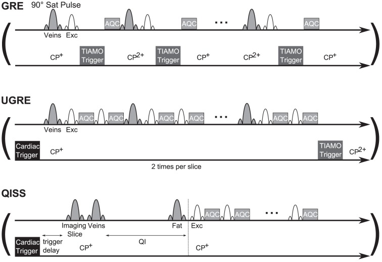
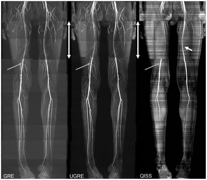
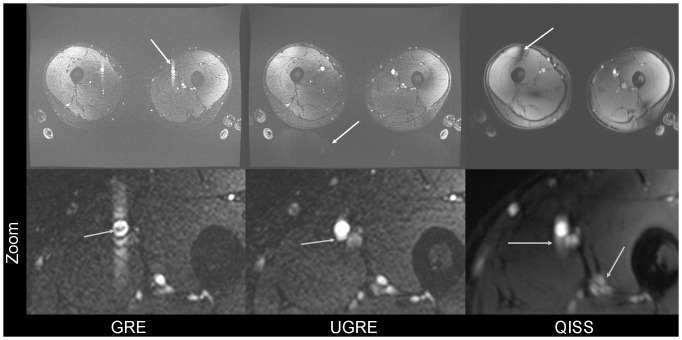
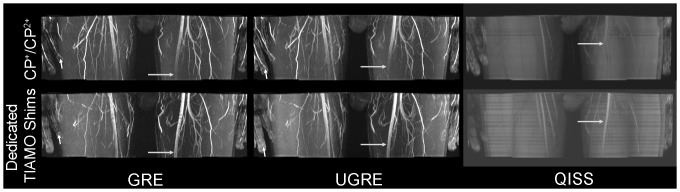
In this study three sequences for non-contrast-enhanced MRA of the lower extremity arteries at 7T were compared. Cardiac triggering was used with the aim to reduce signal variations in the arteries. Two fast single-shot 2D sequences, a modified Ultrafast Spoiled Gradient Echo (UGRE) sequence and a variant of the Quiescent-Interval Single-Shot (QISS) sequence were triggered via phonocardiogram and compared in volunteer examinations to a non-triggered 2D gradient echo (GRE) sequence. For image acquisition, a 16-channel transmit/receive coil and a manually positionable AngioSURF table were used. To tackle B1 inhomogeneities at 7T, Time-Interleaved Acquisition of Modes (TIAMO) was integrated in GRE and UGRE. To compare the three sequences quantitatively, a vessel-to-background ratio (VBR) was measured in all volunteers and stations. In conclusion, cardiac triggering was able to suppress flow artifacts satisfactorily. The modified UGRE showed only moderate image artifacts. Averaged over all volunteers and stations, GRE reached a VBR of 4.18±0.05, UGRE 5.20±0.06, and QISS 2.72±0.03. Using cardiac triggering and TIAMO imaging technique was essential to perform non-enhanced MRA of the lower extremities vessels at 7T. The modified UGRE performed best, as observed artifacts were only moderate and the highest average VBR was reached.


Conflict of interest statement

Competing Interests: The authors have declared that no competing interests exist.

Figures

Figure 1
Figure 1. Sequence diagrams of GRE, UGRE and QISS.
In GRE, no cardiac triggering is used. Here, the TIAMO trigger changes the excitation mode directly after every excitation pulse, which means that both modes needed for a complete slice are acquired in an interleaved fashion. Saturation pulses are applied every TR. In UGRE, the cardiac trigger event starts the acquisition of a complete slice in a single shot. Due to TIAMO, this acquisition has to be repeated to acquire the same slice with the second mode. Venous saturation RF pulses are applied only sparsely. QISS uses three different saturation pulses to prepare the image contrast: In the imaging slice to suppress background tissue, a travelling venous saturation pulse below the imaging slice and a fat saturation pulse in the imaging slice. After the venous saturation, a time interval QI allows unsaturated arterial blood spins to enter the imaging slice. No saturation pulses are applied during the single-shot slice acquisition. The duration of the trigger events is shorter than pictured in these diagrams.
Figure 2
Figure 2. Complete MIPs of GRE, UGRE and QISS of one volunteer.
MIPs of all acquired stations merged together manually in one volunteer for GRE, UGRE, and QISS. In this volunteer, the two stations covering the upper part of the thighs (marked by white double-headed arrows) used individual RF shims to prevent complete artery signal dropout. Vessel signal fluctuation in GRE could be mostly avoided in UGRE and QISS due to cardiac triggering (grey arrows). Tissue signal intensity fluctuation in QISS is most probably due to time delay induced by heartbeat triggering giving the surrounding tissue time to relax. White arrow in QISS shows vein visible in QISS sequence, as only one of the TIAMO RF shim modes could be used, preventing complete venous suppression.
Figure 3
Figure 3. Signal variation flow artifact.
Signal variation flow artifact (white arrow in GRE) shown for consecutive axial slices at position of the grey arrow in Fig. 1 compared to the corresponding images in UGRE and QISS. In UGRE large arteries seem to be slightly broadened (white arrow). In the QISS images, B1 inhomogeneities are considerably more pronounced as TIAMO could not be used (white arrows). For GRE and UGRE, especially the smaller vessels can be more easily delineated than for QISS (grey arrows). The upper row shows the slice position superior to the images of the lower row.
Figure 4
Figure 4. Artifacts in GRE, UGRE and QISS.
The upper row visualizes the flow artifact in GRE images, aliasing in UGRE, and B1 inhomogeneity in QISS, in each case marked by a white arrow. Due to use of TIAMO, the B1 inhomogeneity could be ameliorated in GRE and UGRE. Zoomed images of the left thigh in the lower row show GRE, where the flow artifact in the phase-encode direction and a small signal dropout inside the vessel lumen are clearly visible (grey arrow), UGRE and QISS. For UGRE, slight broadening of the arteries can be observed (grey arrow). In the QISS image, blurring of the vessel walls is clearly observed (grey arrows). Artery circumference can be delineated best in UGRE.
Figure 5
Figure 5. Dedicated TIAMO shims.
MIPs showing the first station of the thigh. The upper row shows images acquired using TIAMO with the CP+ and CP2+ modes. In the case of QISS, only CP+ was used. In the lower row, dedicated TIAMO shims were calculated after the acquisition of B1 maps. In the case of QISS, only one of the two modes could be used. One can clearly see that the left superficial femoral artery can be delineated throughout the entire station with the dedicated B1 shims (grey arrows), whereas the finger arteries lost signal (short white arrow in GRE and UGRE).

Similar articles

Cited by

References

    1. Gutzeit A, Sutter R, Froehlich JM, Roos JE, Sautter T, et al. (2011) ECG-triggered non-contrast-enhanced MR angiography (TRANCE) versus digital subtraction angiography (DSA) in patients with peripheral arterial occlusive disease of the lower extremities. Eur Radiol 21: 1979–1987. - PubMed
    1. Thomsen HS (2006) Nephrogenic systemic fibrosis: A serious late adverse reaction to gadodiamide. Eur Radiol 16: 2619–2621. - PMC - PubMed
    1. Prince MR, Zhang HL, Roditi GH, Leiner T, Kucharczyk W (2009) Risk factors for NSF: a literature review. J Magn Reson Imaging 30: 1298–1308. - PubMed
    1. Lanzman RS, Blondin D, Schmitt P, Orzechowski D, Godehardt E, et al. (2010) Non-Enhanced 3D MR Angiography of the Lower Extremity using ECG-Gated TSE Imaging with Non-Selective Refocusing Pulses - Initial Experience. Rofo 182: 861–867. - PubMed
    1. Hodnett PA, Koktzoglou I, Davarpanah AH, Scanlon TG, Collins JD, et al. (2011) Evaluation of Peripheral Arterial Disease with Nonenhanced Quiescent-Interval Single-Shot MR Angiography. Radiology 260: 282–293. - PMC - PubMed

Publication types

LinkOut - more resources