. 2013:2013:367206.
doi: 10.1155/2013/367206. Epub 2013 Dec 24.

PDGF suppresses oxidative stress induced Ca2+ overload and calpain activation in neurons

Affiliations

PDGF suppresses oxidative stress induced Ca2+ overload and calpain activation in neurons

Lian-Shun Zheng et al. Oxid Med Cell Longev. 2013.

Abstract

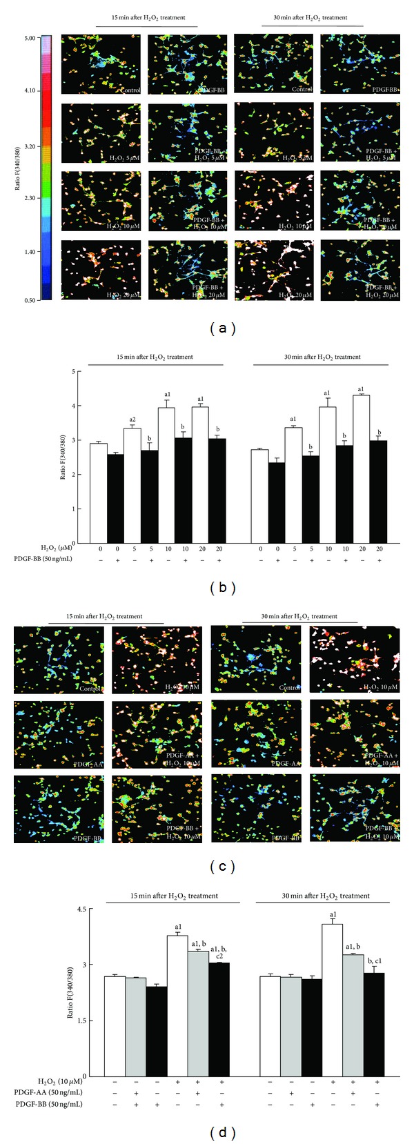
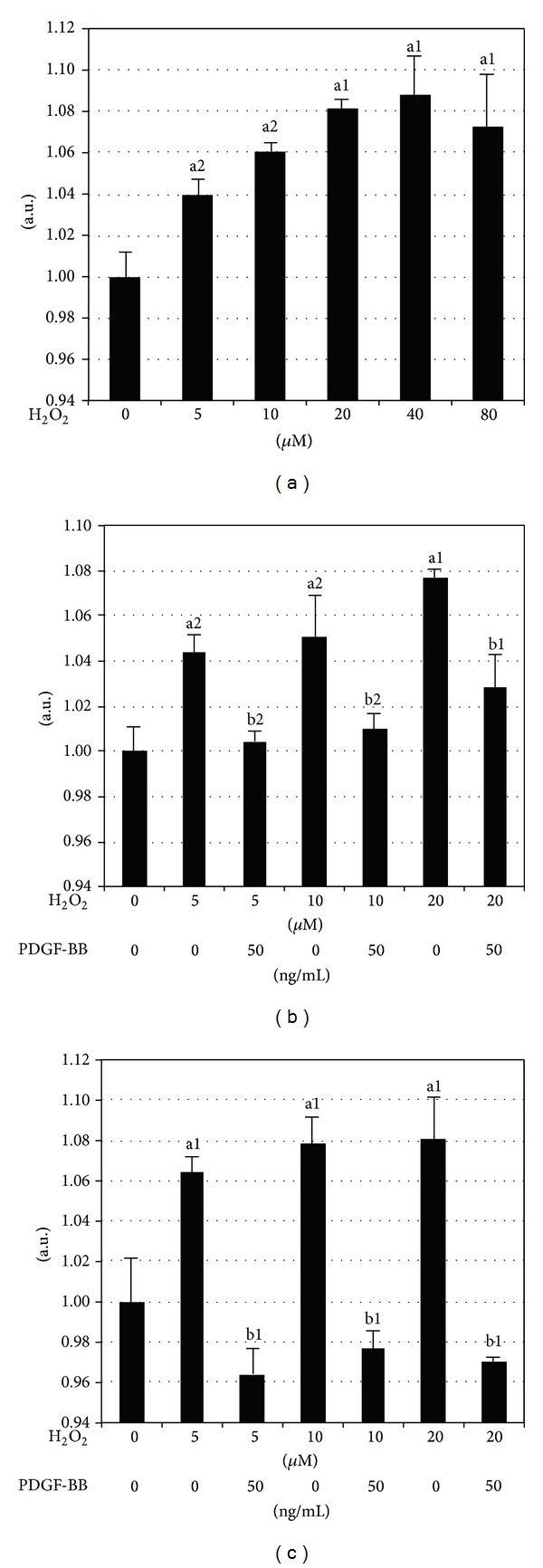
Oxidative stress is crucially involved in the pathogenesis of neurological diseases such as stroke and degenerative diseases. We previously demonstrated that platelet-derived growth factors (PDGFs) protected neurons from H2O2-induced oxidative stress and indicated the involvement of PI3K-Akt and MAP kinases as an underlying mechanism. Ca(2+) overload has been shown to mediate the neurotoxic effects of oxidative stress and excitotoxicity. We examined the effects of PDGFs on H2O2-induced Ca(2+) overload in primary cultured neurons to further clarify their neuroprotective mechanism. H2O2-induced Ca(2+) overload in neurons in a dose-dependent manner, while pretreating neurons with PDGF-BB for 24 hours largely suppressed it. In a comparative study, the suppressive effects of PDGF-BB were more potent than those of PDGF-AA. We then evaluated calpain activation, which was induced by Ca(2+) overload and mediated both apoptotic and nonapoptotic cell death. H2O2-induced calpain activation in neurons in a dose-dependent manner. Pretreatment of PDGF-BB completely blocked H2O2-induced calpain activation. To the best of our knowledge, the present study is the first to demonstrate the mechanism underlying the neuroprotective effects of PDGF against oxidative stress via the suppression of Ca(2+) overload and inactivation of calpain and suggests that PDGF-BB may be a potential therapeutic target of neurological diseases.


Figures

Figure 1
Figure 1
Effects of PDGF-AA and PDGF-BB on the H2O2-induced increase in [Ca2+]i. [Ca2+]i was determined using Fura-2-AM fluorescent dye. (a) Pseudocolor images representing the relative [Ca2+]i indicated by the fluorescence ratio between 340 and 380 nm (F340/380) in individual cortical neurons 15 and 30 min after the H2O2 treatment. Following a 24 h preincubation with 50 ng/mL PDGF-BB, neurons were loaded with Fura-2-AM and exposed to different concentrations of H2O2 (5, 10, and 20 μM). The inserted bar indicates the relationship between colors and fluorescent intensity ratios at 340 and 380 nm. (b) Histogram analyses of the mean F340/380 of (a). Three pictures were taken from each well, and the means of the F340/380 of each neuron were calculated. Data are expressed as means ± SEM derived from three different sets of experiments. (c) To compare the effects of PDGF-AA and PDGF-BB on [Ca2+]i overload induced by H2O2, cells were pretreated with 50 ng/mL PDGF-AA or PDGF-BB for 24 h followed by exposure to 10 μM H2O2 for 15 and 30 min. (d) Histogram analysis of (c). Data are expressed as means ± SEM of three independent experiments. a1 P < 0.01 and a2 P < 0.05 versus the untreated control; b P < 0.01 versus the same H2O2 exposure without the PDGF pretreatment; c1 P < 0.01 and c2 P < 0.05 versus the same H2O2 exposure with the PDGF-AA pretreatment.
Figure 2
Figure 2
Effects of PDGF-BB on H2O2-induced calpain activation. Calpain-1 activity was measured 24 h after exposure to H2O2 as described in the Experimental procedures. (a) Calpain activity induced by the indicated amounts of H2O2 (5, 10, 20, 40, and 80 μM) exposure increased calpain activation in cortical neurons in a dose-dependent manner from 5–20 μM. ((b), (c)) Calpain activity in neuronal cultures pretreated with 50 ng/mL PDGF-BB 24 h (b) and 48 h (c) before exposure to H2O2 PDGF-BB completely blocked calpain activity induced by H2O2. Data are expressed as means ± SEM of three independent experiments. a1 P < 0.01 and a2 P < 0.05 versus the untreated control; b1 P < 0.01 and b2 P < 0.05 versus the same H2O2 exposure without the PDGF-BB pretreatment.

Similar articles

Cited by

References

    1. Sayre LM, Perry G, Smith MA. Oxidative stress and neurotoxicity. Chemical Research in Toxicology. 2008;21(1):172–188. - PubMed
    1. Chan PH, Niizuma K, Endo H. Oxidative stress and mitochondrial dysfunction as determinants of ischemic neuronal death and survival. Journal of Neurochemistry. 2009;109(1):133–138. - PMC - PubMed
    1. Gandhi S, Abramov AY. Mechanism of oxidative stress in neurodegeneration. Oxidative Medicine and Cellular Longevity. 2012;2012:11 pages.428010 - PMC - PubMed
    1. Krieger C, Duchen MR. Mitochondria, Ca2+ and neurodegenerative disease. European Journal of Pharmacology. 2002;447(2-3):177–188. - PubMed
    1. Orrenius S, Zhivotovsky B, Nicotera P. Regulation of cell death: the calcium-apoptosis link. Nature Reviews Molecular Cell Biology. 2003;4(7):552–565. - PubMed

Publication types

LinkOut - more resources