

Sinus lift and transantral approach to root fragment removal
- PMID: 24455319
- PMCID: PMC3886442
- DOI: 10.1155/2013/612108
Sinus lift and transantral approach to root fragment removal
Abstract
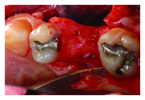
The aim of this case report is to present a case of root fragment removal during planned sinus lift procedure. After failed molar tooth extraction, we chose to retrieve the residual root apex with transantral approach not to damage excessively bone volume. Without changing primary implant rehabilitation purpose, the fragment removal procedure was performed prior to implant placement during necessary sinus lift surgery. Higher visibility of surgical field was achieved. The root fragment residual was removed without an additional surgery appointment avoiding postoperative discomfort. The goal is to underline the importance of being able to change planning during intrasurgical complications. It is most appropriate to operate with safe and simple procedures to reduce surgical discomfort for the patient.
Figures


Similar articles
-
Palatal root surgery of a maxillary molar using a piezosurgery transantral approach with simultaneous sinus lift grafting: a case report.Int Endod J. 2021 Mar;54(3):464-475. doi: 10.1111/iej.13423. Epub 2020 Oct 29. Int Endod J. 2021. PMID: 33012051
-
1-stage versus 2-stage lateral sinus lift procedures: 1-year post-loading results of a multicentre randomised controlled trial.Eur J Oral Implantol. 2014 Spring;7(1):65-75. Eur J Oral Implantol. 2014. PMID: 24892114 Clinical Trial.
-
1-stage versus 2-stage lateral maxillary sinus lift procedures: 4-month post-loading results of a multicenter randomised controlled trial.Eur J Oral Implantol. 2013 Summer;6(2):153-65. Eur J Oral Implantol. 2013. PMID: 23926587 Clinical Trial.
-
Recent Trends in Sinus Lift Surgery and Their Clinical Implications.Clin Implant Dent Relat Res. 2016 Feb;18(1):204-12. doi: 10.1111/cid.12275. Epub 2014 Oct 2. Clin Implant Dent Relat Res. 2016. PMID: 25274014 Review.
-
Implants in the Posterior Maxilla: Open Sinus Lift Versus Conventional Implant Placement. A Systematic Review.Int J Oral Maxillofac Implants. 2019 July/August;34(4):e65–e76. doi: 10.11607/jomi.7274. Epub 2019 Feb 26. Int J Oral Maxillofac Implants. 2019. PMID: 30807622
References
-
- Bouchard P, Renouard F, Bourgeois D, Fromentin O, Jeanneret MH, Beresniak A. Cost-effectiveness modeling of dental implant vs. bridge. Clinical Oral Implants Research. 2009;20(6):583–587. - PubMed
-
- Adell R, Lekholm U, Rockler B, Branemark PI. A 15-year study of osseointegrated implants in the treatment of the edentulous jaw. International Journal of Oral Surgery. 1981;10(6):387–416. - PubMed
-
- Venkateshwar G, Padhye M, Khosla A, Kakkar S. Complications of exodontia: a retrospective study. Indian Journal of Dental Research. 2011;22(5):633–638. - PubMed
-
- Uckan S, Buchbinder D. Sinus lift approach for the retrieval of root fragments from the maxillary sinus. International Journal of Oral and Maxillofacial Surgery. 2003;32(1):87–90. - PubMed
LinkOut - more resources
Full Text Sources
Other Literature Sources
Miscellaneous
