

Ultrasound prenatal diagnosis of inguinal scrotal hernia and contralateral hydrocele
- PMID: 24455356
- PMCID: PMC3876836
- DOI: 10.1155/2013/764579
Ultrasound prenatal diagnosis of inguinal scrotal hernia and contralateral hydrocele
Abstract
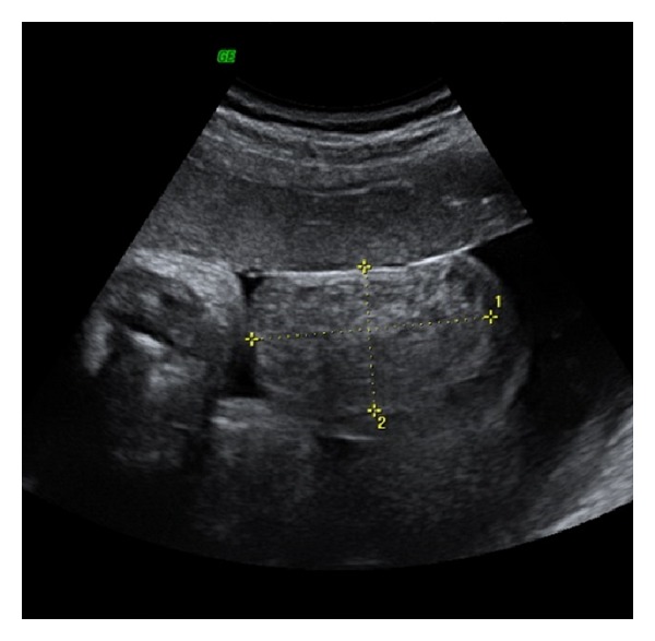
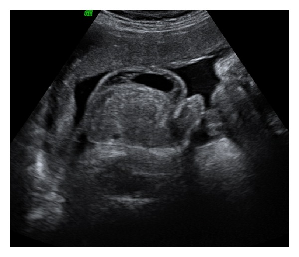
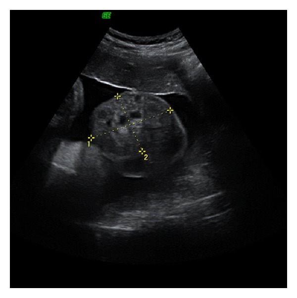
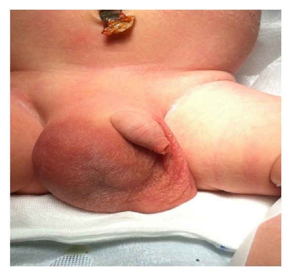
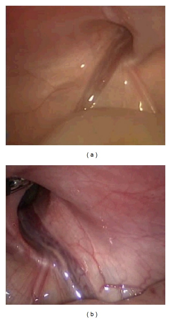
Fetal inguinal scrotal hernia is a rare condition resulting in an abnormal embryonic process of the tunica vaginalis. We report a case of ultrasound prenatal diagnosis of inguinal scrotal hernia associated with contralateral hydrocele in a woman at 37 weeks of gestation, referred to our clinic for a scrotal mass. Differential diagnosis includes hydrocele, teratoma, hemangiomas, solid tumours of testis, bowel herniation, and testicular torsion. Bowel peristalsis is an important ultrasound sign and it allowed us to make diagnosis of inguinal scrotal hernia. Diagnosis was confirmed at birth and a laparoscopic hernia repair was performed without complications on day 10. During surgery, a bilateral defect of canal inguinal was seen and considered as the cause of scrotal inguinal hernia and contralateral hydrocele observed in utero.
Figures
Similar articles
-
Treatment of contralateral hydrocele in neonatal testicular torsion: Is less more?J Pediatr Urol. 2016 Oct;12(5):306.e1-306.e4. doi: 10.1016/j.jpurol.2015.07.009. Epub 2015 Sep 5. J Pediatr Urol. 2016. PMID: 26708803
-
Prenatal diagnosis of scrotal-inguinal hernia: two case reports and review of the English literature.Eur J Obstet Gynecol Reprod Biol. 2013 Nov;171(1):9-11. doi: 10.1016/j.ejogrb.2013.07.026. Epub 2013 Aug 2. Eur J Obstet Gynecol Reprod Biol. 2013. PMID: 23916582 Review.
-
The very large recurrent postoperative scrotal hydrocele after pediatric inguinal hernia repair: a rare problem.Pediatr Surg Int. 2009 Mar;25(3):239-41. doi: 10.1007/s00383-009-2326-x. Epub 2009 Jan 30. Pediatr Surg Int. 2009. PMID: 19184055
-
Ascending testis after repair of pediatric inguinal hernia and hydrocele: A misunderstood operative complication.J Pediatr Urol. 2017 Feb;13(1):53.e1-53.e5. doi: 10.1016/j.jpurol.2016.08.013. Epub 2016 Sep 28. J Pediatr Urol. 2017. PMID: 27727095
-
The genitofemoral nerve may link testicular inguinoscrotal descent with congenital inguinal hernia.Aust N Z J Surg. 1996 Sep;66(9):612-7. doi: 10.1111/j.1445-2197.1996.tb00831.x. Aust N Z J Surg. 1996. PMID: 8859162 Review.
Cited by
-
Inguinoscrotal Hernia, a Possible Cause of Rapidly Developing Fetal Scrotal Mass: Case Report and Literature Update.Healthcare (Basel). 2024 Mar 2;12(5):583. doi: 10.3390/healthcare12050583. Healthcare (Basel). 2024. PMID: 38470694 Free PMC article.
-
Foetal inguinoscrotal hernia-its prenatal diagnosis and its spontaneous regression.BJR Case Rep. 2016 Feb 18;2(2):20150277. doi: 10.1259/bjrcr.20150277. eCollection 2016. BJR Case Rep. 2016. PMID: 30363624 Free PMC article.
-
Sonographic evaluation of fetal scrotum, testes and epididymis.Obstet Gynecol Sci. 2021 Sep;64(5):393-406. doi: 10.5468/ogs.21040. Epub 2021 Jun 28. Obstet Gynecol Sci. 2021. PMID: 34176256 Free PMC article.
-
Classifying Hydroceles of the Pelvis and Groin: An Overview of Etiology, Secondary Complications, Evaluation, and Management.Curr Urol. 2017 Apr;10(1):1-14. doi: 10.1159/000447145. Epub 2017 Mar 30. Curr Urol. 2017. PMID: 28559772 Free PMC article. Review.
References
-
- Skoog SJ, Conlin MJ. Pediatric hernias and hydroceles: the urologist’s perspective. Urologic Clinics of North America. 1995;22(1):119–130. - PubMed
-
- Shadbolt CL, Heinze SBJ, Dietrich RB. Imaging of groin masses: inguinal anatomy and pathologic conditions revisited. Radiographics. 2001;21:S261–S271. - PubMed
-
- Rowe MI, Copelson LW, Clatworthy HW., Jr. The patent processus vaginalis and the inguinal hernia. Journal of Pediatric Surgery. 1969;4(1):102–107. - PubMed
-
- Ober KJ, Smith CV. Prenatal ultrasound diagnosis of a fetal inguinal hernia containing small bowel. Obstetrics and Gynecology. 1991;78(5):905–906. - PubMed
-
- Meizner I, Levy A, Katz M, Simhon T, Glezerman M. Prenatal ultrasonographic diagnosis of fetal scrotal inguinal hernia. American Journal of Obstetrics and Gynecology. 1992;166(3):907–909. - PubMed
LinkOut - more resources
Full Text Sources
Other Literature Sources
