

Ultrasound prenatal diagnosis of inguinal scrotal hernia and contralateral hydrocele
- PMID: 24455356
- PMCID: PMC3876836
- DOI: 10.1155/2013/764579
Ultrasound prenatal diagnosis of inguinal scrotal hernia and contralateral hydrocele
Abstract
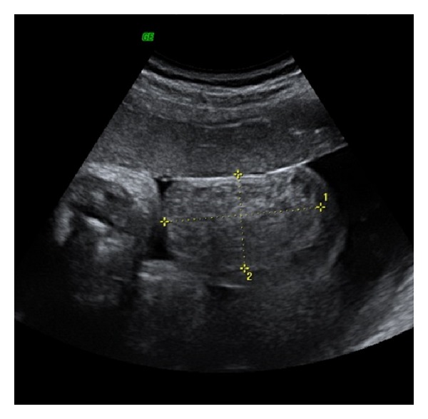
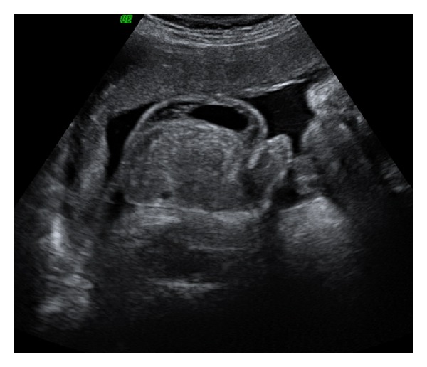
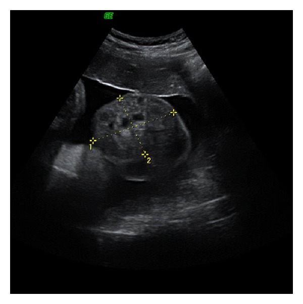
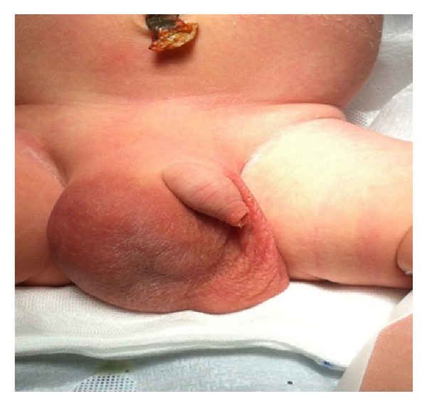
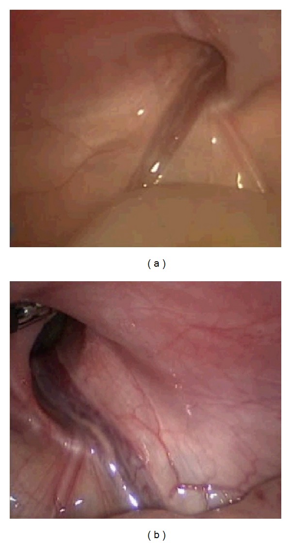
Fetal inguinal scrotal hernia is a rare condition resulting in an abnormal embryonic process of the tunica vaginalis. We report a case of ultrasound prenatal diagnosis of inguinal scrotal hernia associated with contralateral hydrocele in a woman at 37 weeks of gestation, referred to our clinic for a scrotal mass. Differential diagnosis includes hydrocele, teratoma, hemangiomas, solid tumours of testis, bowel herniation, and testicular torsion. Bowel peristalsis is an important ultrasound sign and it allowed us to make diagnosis of inguinal scrotal hernia. Diagnosis was confirmed at birth and a laparoscopic hernia repair was performed without complications on day 10. During surgery, a bilateral defect of canal inguinal was seen and considered as the cause of scrotal inguinal hernia and contralateral hydrocele observed in utero.
Figures
References
-
- Skoog SJ, Conlin MJ. Pediatric hernias and hydroceles: the urologist’s perspective. Urologic Clinics of North America. 1995;22(1):119–130. - PubMed
-
- Shadbolt CL, Heinze SBJ, Dietrich RB. Imaging of groin masses: inguinal anatomy and pathologic conditions revisited. Radiographics. 2001;21:S261–S271. - PubMed
-
- Rowe MI, Copelson LW, Clatworthy HW., Jr. The patent processus vaginalis and the inguinal hernia. Journal of Pediatric Surgery. 1969;4(1):102–107. - PubMed
-
- Ober KJ, Smith CV. Prenatal ultrasound diagnosis of a fetal inguinal hernia containing small bowel. Obstetrics and Gynecology. 1991;78(5):905–906. - PubMed
-
- Meizner I, Levy A, Katz M, Simhon T, Glezerman M. Prenatal ultrasonographic diagnosis of fetal scrotal inguinal hernia. American Journal of Obstetrics and Gynecology. 1992;166(3):907–909. - PubMed
LinkOut - more resources
Full Text Sources
Other Literature Sources
