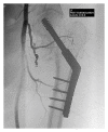
Arterial Injury to the Profunda Femoris Artery following Internal Fixation of a Neck of Femur Fracture with a Compression Hip Screw
- PMID: 24455367
- PMCID: PMC3877589
- DOI: 10.1155/2013/181293
Arterial Injury to the Profunda Femoris Artery following Internal Fixation of a Neck of Femur Fracture with a Compression Hip Screw
Abstract
We report the case of an 82-year-old woman who developed extensive proximal thigh swelling and persistent anaemia following internal fixation of an extracapsular neck of femur fracture with a dynamic hip screw (DHS). This was revealed to be a pseudoaneurysm of a branch of profunda femoris artery on angiography. Her case was further complicated by a concurrent pulmonary embolism (PE). She underwent endovascular coil embolisation of the pseudoaneurysm. An IVC filter was inserted and the patient was fully anticoagulated once it had been ensured that there was no active bleeding. In this case, we review the potential for anatomical variations in the blood supply to this region and discuss treatment options for a complicated patient. We recommend that a pseudoaneurysm should be part of a differential diagnosis for postoperative patients with anaemia refractory to blood transfusion so as not to miss this rare but potentially serious complication.
Figures


References
-
- Barnes DI, Broude HB. False aneurysm of the profunda femoris artery complicating fracture of the femoral shaft and treated by transcatheter embolization. A case report. South African Medical Journal. 1985;67(20):824–826. - PubMed
-
- Canbaz S, Acipayam M, Gürbüz H, Duran E. False aneurysm of perforating branch of the profunda femoris artery after external fixation for a complicated femur fracture. Journal of Cardiovascular Surgery. 2002;43(4):519–521. - PubMed
-
- Jacobs E, Groot D, Das M, Hermus JPS. Pseudoaneurysm of the anterior tibial artery after ankle arthroscopy. Journal of Foot and Ankle Surgery. 2011;50(3):361–363. - PubMed
LinkOut - more resources
Full Text Sources
Other Literature Sources

