

Protein Machineries Involved in the Attachment of Heme to Cytochrome c: Protein Structures and Molecular Mechanisms
- PMID: 24455431
- PMCID: PMC3884852
- DOI: 10.1155/2013/505714
Protein Machineries Involved in the Attachment of Heme to Cytochrome c: Protein Structures and Molecular Mechanisms
Abstract
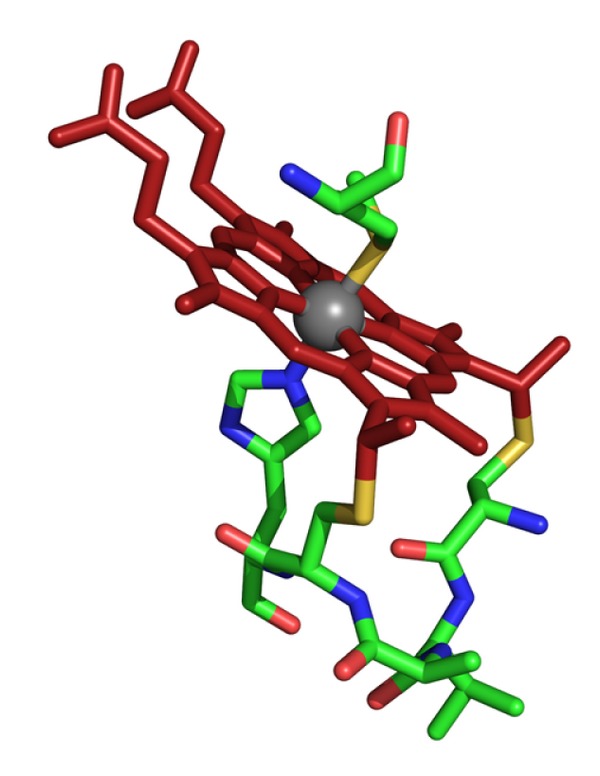
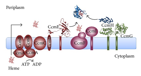
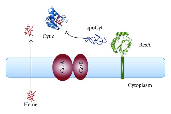
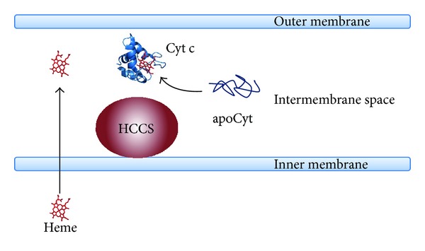
Cytochromes c (Cyt c) are ubiquitous heme-containing proteins, mainly involved in electron transfer processes, whose structure and functions have been and still are intensely studied. Surprisingly, our understanding of the molecular mechanism whereby the heme group is covalently attached to the apoprotein (apoCyt) in the cell is still largely unknown. This posttranslational process, known as Cyt c biogenesis or Cyt c maturation, ensures the stereospecific formation of the thioether bonds between the heme vinyl groups and the cysteine thiols of the apoCyt heme binding motif. To accomplish this task, prokaryotic and eukaryotic cells have evolved distinctive protein machineries composed of different proteins. In this review, the structural and functional properties of the main maturation apparatuses found in gram-negative and gram-positive bacteria and in the mitochondria of eukaryotic cells will be presented, dissecting the Cyt c maturation process into three functional steps: (i) heme translocation and delivery, (ii) apoCyt thioreductive pathway, and (iii) apoCyt chaperoning and heme ligation. Moreover, current hypotheses and open questions about the molecular mechanisms of each of the three steps will be discussed, with special attention to System I, the maturation apparatus found in gram-negative bacteria.
Figures
Similar articles
-
Recognition and binding of apocytochrome c to P. aeruginosa CcmI, a component of cytochrome c maturation machinery.Biochim Biophys Acta. 2013 Aug;1834(8):1554-61. doi: 10.1016/j.bbapap.2013.04.027. Epub 2013 May 3. Biochim Biophys Acta. 2013. PMID: 23648553
-
Structural Insights into Mechanisms Underlying Mitochondrial and Bacterial Cytochrome c Synthases.Biomolecules. 2024 Nov 21;14(12):1483. doi: 10.3390/biom14121483. Biomolecules. 2024. PMID: 39766190 Free PMC article.
-
Mechanisms and Control of Heme Transport and Incorporation into Cytochrome c.Annu Rev Microbiol. 2025 Jun 3. doi: 10.1146/annurev-micro-050624-031631. Online ahead of print. Annu Rev Microbiol. 2025. PMID: 40460019 Review.
-
Structure of a trypanosomatid mitochondrial cytochrome c with heme attached via only one thioether bond and implications for the substrate recognition requirements of heme lyase.FEBS J. 2009 May;276(10):2822-32. doi: 10.1111/j.1742-4658.2009.07005.x. FEBS J. 2009. PMID: 19459937
-
Order within a mosaic distribution of mitochondrial c-type cytochrome biogenesis systems?FEBS J. 2008 May;275(10):2385-402. doi: 10.1111/j.1742-4658.2008.06380.x. Epub 2008 Apr 3. FEBS J. 2008. PMID: 18393999 Review.
Cited by
-
A Case of Gene Fragmentation in Plant Mitochondria Fixed by the Selection of a Compensatory Restorer of Fertility-Like PPR Gene.Mol Biol Evol. 2021 Jul 29;38(8):3445-3458. doi: 10.1093/molbev/msab115. Mol Biol Evol. 2021. PMID: 33878189 Free PMC article.
-
Development of a production chain from vegetable biowaste to platform chemicals.Microb Cell Fact. 2018 Jun 13;17(1):90. doi: 10.1186/s12934-018-0937-4. Microb Cell Fact. 2018. PMID: 29898726 Free PMC article.
-
Genome-wide identification of genes probably relevant to the adaptation of schizothoracins (Teleostei: Cypriniformes) to the uplift of the Qinghai-Tibet Plateau.BMC Genomics. 2017 Apr 20;18(1):310. doi: 10.1186/s12864-017-3703-9. BMC Genomics. 2017. PMID: 28427344 Free PMC article.
-
Evaluative profiling of arsenic sensing and regulatory systems in the human microbiome project genomes.Microbiol Insights. 2014 Nov 11;7:25-34. doi: 10.4137/MBI.S18076. eCollection 2014. Microbiol Insights. 2014. PMID: 25452698 Free PMC article.
-
During Cytochrome c Maturation CcmI Chaperones the Class I Apocytochromes until the Formation of Their b-Type Cytochrome Intermediates.J Biol Chem. 2015 Jul 3;290(27):16989-7003. doi: 10.1074/jbc.M115.652818. Epub 2015 May 15. J Biol Chem. 2015. PMID: 25979338 Free PMC article.
References
Publication types
LinkOut - more resources
Full Text Sources
Other Literature Sources
