Review
. 2013 Dec 19:2:123-39.
doi: 10.1016/j.redox.2013.12.011. eCollection 2014.

Redox regulation of mitochondrial function with emphasis on cysteine oxidation reactions

Affiliations
Review

Redox regulation of mitochondrial function with emphasis on cysteine oxidation reactions

Ryan J Mailloux et al. Redox Biol. .

Abstract

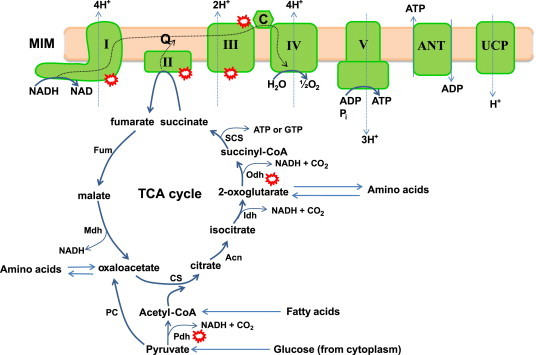
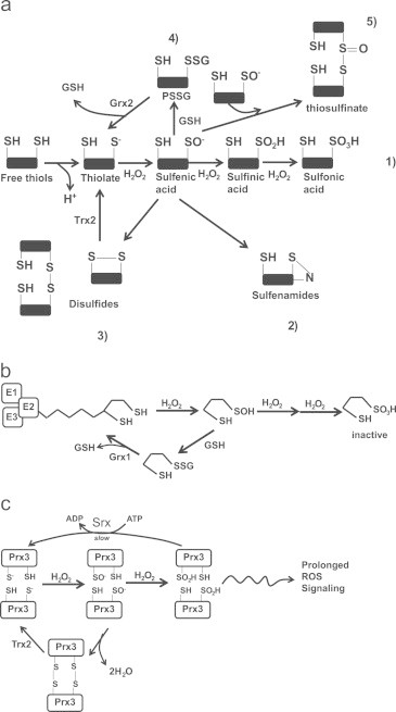
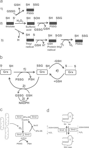
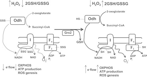
Mitochondria have a myriad of essential functions including metabolism and apoptosis. These chief functions are reliant on electron transfer reactions and the production of ATP and reactive oxygen species (ROS). The production of ATP and ROS are intimately linked to the electron transport chain (ETC). Electrons from nutrients are passed through the ETC via a series of acceptor and donor molecules to the terminal electron acceptor molecular oxygen (O2) which ultimately drives the synthesis of ATP. Electron transfer through the respiratory chain and nutrient oxidation also produces ROS. At high enough concentrations ROS can activate mitochondrial apoptotic machinery which ultimately leads to cell death. However, if maintained at low enough concentrations ROS can serve as important signaling molecules. Various regulatory mechanisms converge upon mitochondria to modulate ATP synthesis and ROS production. Given that mitochondrial function depends on redox reactions, it is important to consider how redox signals modulate mitochondrial processes. Here, we provide the first comprehensive review on how redox signals mediated through cysteine oxidation, namely S-oxidation (sulfenylation, sulfinylation), S-glutathionylation, and S-nitrosylation, regulate key mitochondrial functions including nutrient oxidation, oxidative phosphorylation, ROS production, mitochondrial permeability transition (MPT), apoptosis, and mitochondrial fission and fusion. We also consider the chemistry behind these reactions and how they are modulated in mitochondria. In addition, we also discuss emerging knowledge on disorders and disease states that are associated with deregulated redox signaling in mitochondria and how mitochondria-targeted medicines can be utilized to restore mitochondrial redox signaling.

Keywords: Mitochondria; Redox; S-glutathionylation; S-nitrosylation; S-oxidation.


Figures

Fig. 1
Fig. 1
Aerobic respiration, oxidative phosphorylation, and chemiosmotic theory: nutrients in the form of pyruvate (generated from glucose via glycolysis in the cytoplasm), acetyl-CoA (produced from pyruvate or generated by fatty acid oxidation), or amino acids enter the tricarboxylic acid (TCA) and are systematically oxidized by the concerted action of 8 different enzymes. Carbon oxidation is coupled to decarboxylation reactions which yield the necessary electrons required to drive oxidative phosphorylation (OXPHOS). Electrons from the TCA cycle are transferred to NAD generating the electron carrier NADH which is then oxidized by Complex I. Electrons can also be provided by Complex II which oxidizes succinate to fumarate in the TCA cycle. Electrons then flow through a series of redox active prosthetic groups and molecules to the terminal electron O2 at Complex IV. The favorable energy change associated with electron flow is coupled to the pumping of protons through Complexes I, III, and IV into the intermembrane space. The protons are then transported back into the matrix by ATP synthase which couples the favorable energy change of proton transfer to the production of ATP from ADP and Pi. This is referred to as “coupled” respiration or OXPHOS. ATP is then exported into the cytoplasm to do “work” in the cell. The antiporter ANT is responsible for exporting ATP in exchange for ADP. The potential difference created by nutrient oxidation can be “uncoupled” from OXPHOS by MIM proteins ANT and UCP for the purpose of thermogenesis or controlling mitochondrial ROS production. Dotted line: represents flow of electrons and protons. Red circles: represent major sites for ROS production in mitochondria. Note that the ROS species is not defined in the figure and that other relevant sources of ROS, dihydroorotate dehydrogenase, sn-glycerol-3-phosphate dehydrogenase (mGPDH), electron transfer flavoprotein:ubiquinol oxidoreductase, p66shc/Cytochrome C, Mia40p/Erv1p, have been excluded for simplicity. Pdh; pyruvate dehydrogenase, PC; pyruvate carboxylase, CS; citrate synthase, Acn; aconitase, Idh; isocitrate dehydrogenase, Odh; 2-oxoglutarate dehydrogenase, SCS; succinyl-CoA synthase, Fum; fumarase, Mdh; malate dehydrogenase, I; Complex I, II; Complex II, III; Complex III, IV; Complex IV, V; Complex V, ANT; adenine nucleotide translocase, UCP; uncoupling protein, Q; quinone, C; cytochrome C.
Fig. 2
Fig. 2
S-oxidation reactions and H2O2 signaling: (a) the various thiol oxidation reactions. Thiols are deprotonated generating a reactive thiolate anion which can then be oxidized by H2O2 to produce a SO. Due to their highly reactive nature, SO undergoes a range of other reactions. Depending on the conditions, environment, and proximity of the SO to other reactive groups SO can be (1) further oxidized by H2O2 to SO2H and SO3H, (2) react with amides on peptide backbones to generate sulfenamides, (3) cross react with neighboring thiols to form intra or intermolecular disulfide bonds which are reversed by Trx2, (4) undergo S-glutathionylation which is reversed by Grx2, and (5) react with a sulfenic acid to generate a thiosulfinate. (b) Lipoic acid residue on the E2 subunit of Odh can be protected from further oxidation by S-glutathionylation. H2O2 oxidizes a sulfhydryl moiety on the lipoate molecule generating SO which can be oxidized further rendering Odh inactive. To protect from further oxidation, the SO is S-glutathionylated which is reversed by Grx to regenerate an active enzyme. (c) Prx3 can adopt multiple oxidation states. The peroxidatic Cys of Prx3 is oxidized by H2O2 to yield SO which then reacts with a neighboring resolving Cys to produce a disulfide bridge. The Cys residues are then reduced by Trx2 to regenerate active Prx3. SO can also be further oxidized to SO2H which maintains Prx3 in an inactive state for a longer period which allows H2O2 signaling to persist. Prx3 is reduced back to an active enzyme complex by Srx. H2O2; hydrogen peroxide, SO; sulfenic acid, SO2H; sulfinic acid, SO3H sulfonic acid, PSSG; protein glutathione disulfide mixture, Odh; 2-oxoglutarate dehydrogenase, Grx; glutaredoxin, Prx3; peroxiredoxin-3, Trx2; thioredoxin-2.
Fig. 3
Fig. 3
Non-enzymatic and enzymatic S-glutathionylation reactions and the modulation of Grx2. (a) Non-enzymatic glutathionylation reactions. (1) Solvent-exposed thiolate is S-glutathionylated by GSSG via a thiol disulfide exchange reaction. (2) Solvent-exposed thiolate are oxidized to a sulfenic acid residue by H2O2 which then undergoes S-glutathionylation. (3) one electron reduction of a thiol forms a thiyl radical which interacts with glutathione to form a glutathione anion thiyl radical intermediate. A disulfide bond then forms between the glutathione anion and the thiyl radical following the one electron reduction of O2 to O2.(b) Catalytic cycle of Grx1 and Grx2 (in diagram it is simply referred to as Grx). In step 1 Grx catalyzes the rapid transfer of the gluthionyl moiety via a disulfide exchange reaction to its catalytic cysteine which produces a Grx-SSG intermediate and a deglutathionylated protein. Step 2 involves the GSH-mediated removal of the glutathionyl moiety from Grx-SSG which generates a fully reduced Grx enzyme and GSSG. For step 3 the GSSG is reduced by NADPH and glutathione reductase to regenerate GSH. Note the side reaction for Grx-SSG in step 4 an intramolecular disulfide can form in Grx. GSH is required to return Grx-SSG to its catalytic cycle. (c) Grx2 is modulated by 2Fe–2S cluster coordination and O2 mediated dissembly of the cluster. Grx2 is maintained is inactive as a homodimer. A subsequent burst of O2 results in the release of active Grx2 monomers which subsequently deglutathionylate target proteins. (d) Grx2 catalyzes the reversible S-glutathionylation of Complex I. When the 2GSH/GSSG ratio is low and H2O2 is high, Grx2 glutathionylase is activated. A high 2GSH/GSSG ratio and low H2O2 levels induce Grx2-mediated deglutathionylation of Complex I.
Fig. 4
Fig. 4
Hypothetical scheme depicting the regulation of mitochondrial O2 production and OXPHOS by S-glutathionylation. When H2O2 is high and 2GSH/GSSG is low in mitochondria, Odh, Complex I, and ATP synthase are temporarily S-glutathionylated to diminish NADH production and limit electron flow through the respiratory chain. Although this may diminish mitochondrial ATP production S-glutathionylation will limit O2 and H2O2 production by mitochondria. In addition, S-glutathionylation of ATP synthase may also prevent ATP hydrolysis and the pumping of protons back to the intermembrane space. This would effectively maintain a lower membrane potential and limit ROS genesis. Once ROS levels have decreased and the 2GSH/GSSG ratio is restored Grx2 deglutathionylates Complex I and potentially Odh and ATP synthase which restores mitochondrial ATP production.

References

    1. Brown G.C., Borutaite V. There is no evidence that mitochondria are the main source of reactive oxygen species in mammalian cells. Mitochondrion. 2012;12:1–4. - PubMed
    1. Fernie A.R., Carrari F., Sweetlove L.J. Respiratory metabolism: glycolysis, the TCA cycle and mitochondrial electron transport. Curr. Opin. Plant Biol. 2004;7:254–261. - PubMed
    1. Baradaran R., Berrisford J.M., Minhas G.S., Sazanov L.A. Crystal structure of the entire respiratory complex I. Nature. 2013;494:443–448. - PMC - PubMed
    1. Quinlan C.L., Orr A.L., Perevoshchikova I.V., Treberg J.R., Ackrell B.A., Brand M.D. Mitochondrial complex II can generate reactive oxygen species at high rates in both the forward and reverse reactions. J. Biol. Chem. 2012;287:27255–27264. - PMC - PubMed
    1. Khutornenko A.A., Roudko V.V., Chernyak B.V., Vartapetian A.B., Chumakov P.M., Evstafieva A.G. Pyrimidine biosynthesis links mitochondrial respiration to the p53 pathway. Proc. Natl. Acad. Sci. USA. 2010;107:12828–12833. - PMC - PubMed

Publication types

LinkOut - more resources