

Squamous cell carcinoma lung: Presented with bilateral lower limb deep venous thrombosis with gangrene formation
- PMID: 24455526
- PMCID: PMC3876636
- DOI: 10.4103/2278-330X.105858
Squamous cell carcinoma lung: Presented with bilateral lower limb deep venous thrombosis with gangrene formation
Abstract
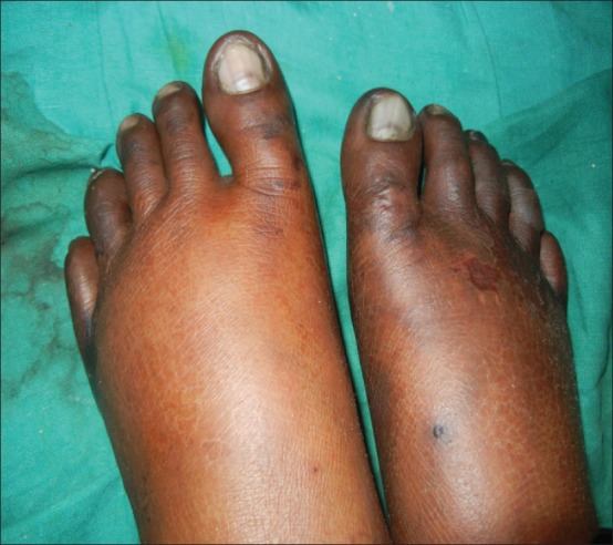
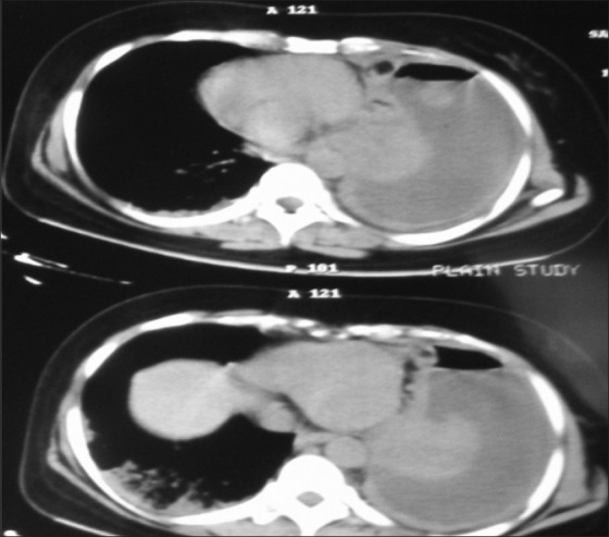
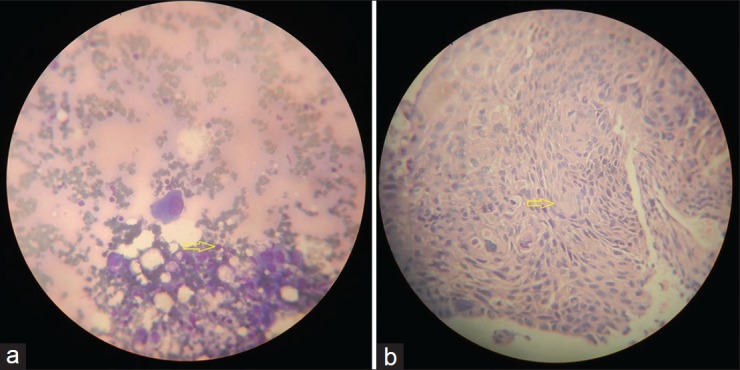
Bilateral venous thrombosis due to underlying malignancy is a rare entity. It is worthy to search for malignancy in patients of bilateral venous gangrene. Our patient presented with severe bilateral leg pain as a result of venous gangrene. There was associated left sided massive pleural effusion with scalp nodule. Fine needle aspiration cytology of scalp nodule revealed metastatic squamous cell carcinoma and fiber optic bronchoscopy guided biopsy from growth at left upper lobe bronchus confirmed the case as squamous cell carcinoma lung. It was rare for squamous cell carcinoma lung to present as bilateral venous gangrene with anticardiolipin antibody negative.
Keywords: Bilateral deep venous thrombosis; squamous cell carcinoma lung; venous gangrene.
Conflict of interest statement
Figures
References
-
- Lee AY, Levine MN. Venous thromboembolism and cancer: Risks and outcomes. Circulation. 2003;107:17–21. - PubMed
-
- Donati MB. Cancer and thrombosis from phlegmasia alba dolens to transgenic mice. Thromb Haemost. 1995;74:278–81. - PubMed
-
- Prandoni P, Falanga A, Piccioli A. Cancer and venous thromboembolism. Lancet Oncol. 2005;6:401–10. - PubMed
-
- Hopper WC, Evatt BL. The role of activated protein C resistance in the pathogenesis of venous thrombosis. Am J Med Sci. 1998;316:120–8. - PubMed
-
- Francis JL, Biggerstaff J, Amirkhosravi A. Hemostasis and malignancy. Semin Thromb Hemost. 1998;24:93–109. - PubMed
Publication types
LinkOut - more resources
Full Text Sources
Other Literature Sources