

Osteosarcoma relapse as pleural metastasis
- PMID: 24455551
- PMCID: PMC3876644
- DOI: 10.4103/2278-330X.110483
Osteosarcoma relapse as pleural metastasis
Abstract
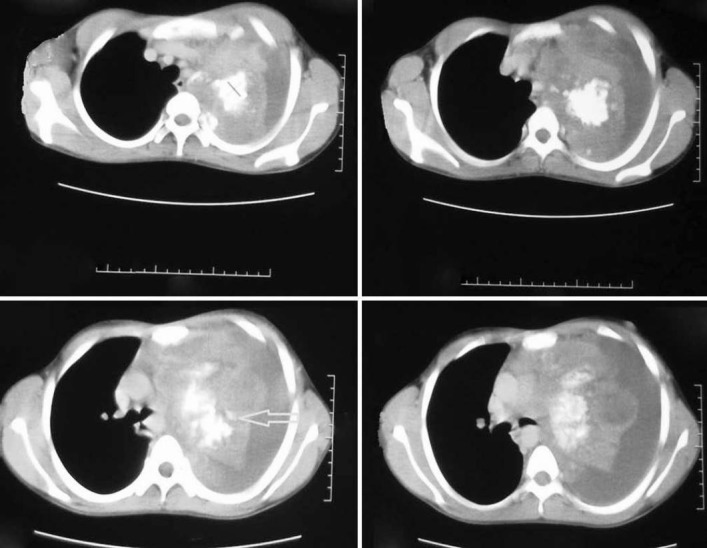
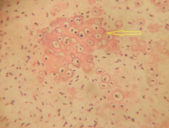
Osteosarcoma is the most common primary bone tumor in children and young adults arising from primitive mesenchymal bone-forming cells. The lung is the most common site of metastasis of osteosarcoma. Here, we report a case of a 14-year-old male patient having osteosarcoma of tibia presenting to us for evaluation of left-sided pleural effusion after 4 years of mid-thigh amputation. Contrast-enhanced computed tomography thorax revealed a large, heterogeneous, calcified mass (+277 H.U) at left upper and middle lobe along with massive left-sided pleural effusion. Thoracoscopy revealed a lung metastasis in the right upper and middle lobe along with 2-cm diameter mass found on the surface of parietal pleura. Lung tumor was resected and biopsy of the pleural mass was carried out. Histopathological examination from both the masses was suggestive of metastatic osteosarcoma. The case underlines the importance of performing thoracoscopy in patients of osteosarcoma who recur with lung metastasis.
Keywords: Metastasis; osteosarcoma; pleura; thoracoscopy.
Conflict of interest statement
Figures
Similar articles
-
Pleural Melanoma Metastasis.Ann Thorac Surg. 2021 Aug;112(2):e103-e105. doi: 10.1016/j.athoracsur.2020.09.088. Epub 2021 Jan 7. Ann Thorac Surg. 2021. PMID: 33421399
-
Extraskeletal osteosarcoma of the pleura: a case report.Gen Thorac Cardiovasc Surg. 2008 Apr;56(4):180-2. doi: 10.1007/s11748-007-0216-5. Epub 2008 Apr 10. Gen Thorac Cardiovasc Surg. 2008. PMID: 18401680
-
PLEURAL METASTASIS OF A PEDIATRIC OSTEOSARCOMA.Port J Card Thorac Vasc Surg. 2022 Jan 4;28(4):55-57. doi: 10.48729/pjctvs.226. Port J Card Thorac Vasc Surg. 2022. PMID: 35334173
-
Left Lung Neoplasms and Bilateral Pleural Effusion Combined Elevated Carcinoembryonic Antigen in Pleural Effusion with Negative Result of Thoracoscopy Pleural Biopsy Misdiagnosed as Lung Carcinoma Ultimately Confirmed Pulmonary Sarcomatoid Carcinoma by CT-guided Percutaneous Lung Biopsy: a Case Report and Literature Review.Clin Lab. 2019 Aug 1;65(8). doi: 10.7754/Clin.Lab.2019.190127. Clin Lab. 2019. PMID: 31414757 Review.
-
Localized Pleural Metastases of Renal Cell Carcinoma After Nephrectomy: A Case Report and Literature Review.J Bronchology Interv Pulmonol. 2016 Jan;23(1):59-62. doi: 10.1097/LBR.0000000000000181. J Bronchology Interv Pulmonol. 2016. PMID: 26705014 Review.
Cited by
-
CRISPR/Cas9: A revolutionary genome editing tool for human cancers treatment.Technol Cancer Res Treat. 2022 Jan-Dec;21:15330338221132078. doi: 10.1177/15330338221132078. Technol Cancer Res Treat. 2022. Retraction in: Technol Cancer Res Treat. 2024 Jan-Dec;23:15330338241240520. doi: 10.1177/15330338241240520. PMID: 36254536 Free PMC article. Retracted. Review.
-
Poly (ADP-ribose) polymerase 1 (PARP1) inhibition promotes pulmonary metastasis of osteosarcoma by boosting ezrin phosphorylation.Int J Biol Sci. 2022 Jan 9;18(3):1238-1253. doi: 10.7150/ijbs.58784. eCollection 2022. Int J Biol Sci. 2022. PMID: 35173550 Free PMC article.
-
Beyond tradition and convention: benefits of non-traditional model organisms in cancer research.Cancer Metastasis Rev. 2021 Mar;40(1):47-69. doi: 10.1007/s10555-020-09930-6. Epub 2020 Oct 28. Cancer Metastasis Rev. 2021. PMID: 33111160 Review.
-
Downregulated lncRNA GAS5 and Upregulated miR-21 Lead to Epithelial-Mesenchymal Transition and Lung Metastasis of Osteosarcomas.Front Cell Dev Biol. 2021 Jul 27;9:707693. doi: 10.3389/fcell.2021.707693. eCollection 2021. Front Cell Dev Biol. 2021. PMID: 34386496 Free PMC article.
-
Osteosarcoma Cell-Derived Exosomal miR-1307 Promotes Tumorgenesis via Targeting AGAP1.Biomed Res Int. 2021 Mar 25;2021:7358153. doi: 10.1155/2021/7358153. eCollection 2021. Biomed Res Int. 2021. PMID: 33834074 Free PMC article.
References
-
- Vander Griend RA. Osteosarcoma and its variants. Orthop Clin North Am. 1996;27:575–81. - PubMed
-
- Tabone MD, Kalifa C, Rodary C, Raquin M, Valteau-Couanet D, Lemerle J. Osteosarcoma recurrences in pediatric patients previously treated with intensive chemotherapy. J Clin Oncol. 1994;12:2614–20. - PubMed
-
- Marina NM, Pratt CB, Rao BN, Shema SJ, Meyer WH. Improved prognosis of children with osteosarcoma metastatic to the lung (s) at the time of diagnosis. Cancer. 1992;70:2722–7. - PubMed
-
- Japan Autopsy Annual Database. Tokyo: Japanese Society of Pathology; 1981-2002. Japanese Society of Pathology. In Japanese.
Publication types
LinkOut - more resources
Full Text Sources
Other Literature Sources
