

A systematic approach to diagnosis of cystic brain lesions
- PMID: 24455569
- PMCID: PMC3876643
- DOI: 10.4103/2278-330X.110509
A systematic approach to diagnosis of cystic brain lesions
Abstract
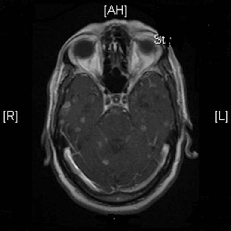
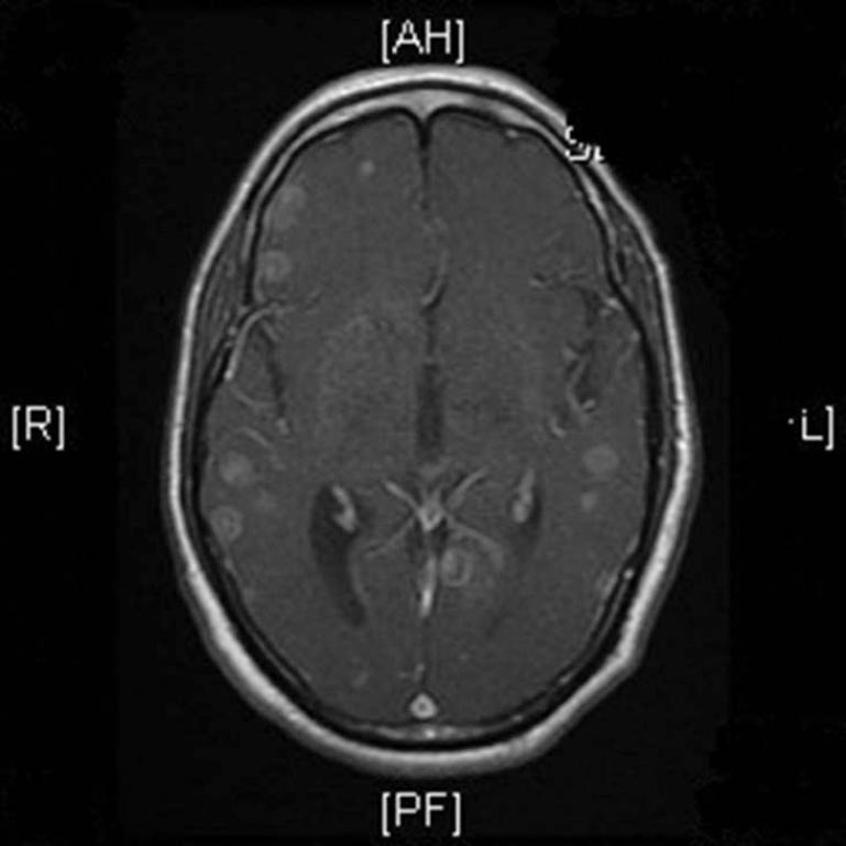
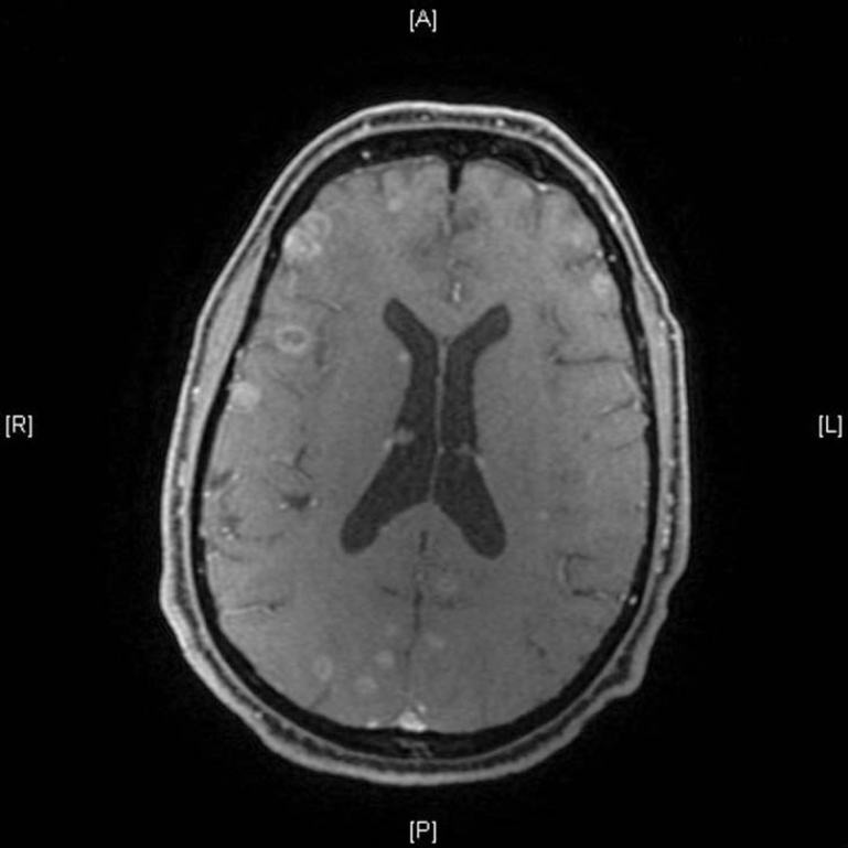
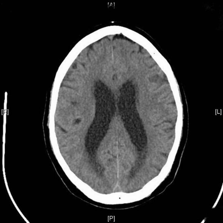
Brain metastasis is the most common intracranial tumor in adults. The incidence of brain metastasis is rising with the increase in survival of cancer patients. Magnetic resonance imaging with contrast enhancement is the imaging procedure of choice to diagnose and characterize brain metastases. Multiple lesions with marked vasogenic edema and mass effect are typically seen in patients with brain metastases. The classical appearance of a metastasis is a solid enhancing mass with well-defined margins and extensive edema. Occasionally, central necrosis produces a ring enhancing mass. Here, we report a case of Non Small Cell Lung Cancer with multiple ring enhancing lesions in brain, and the approach to diagnosis of such patients.
Keywords: Cystic brain metastases; magnetic resonance imaging; neurocysticercosis.
Conflict of interest statement
Figures
Similar articles
-
Pulmonary neuroendocrine carcinoma mimicking neurocysticercosis: a case report.J Med Case Rep. 2016 Jun 2;10(1):144. doi: 10.1186/s13256-016-0910-y. J Med Case Rep. 2016. PMID: 27250121 Free PMC article.
-
Case Report: Atypical Solitary Brain Metastasis: The Role of MR Spectroscopy In Differential Diagnosis.Front Oncol. 2022 Jul 22;12:866622. doi: 10.3389/fonc.2022.866622. eCollection 2022. Front Oncol. 2022. PMID: 35936687 Free PMC article.
-
Brain metastases from adenocarcinoma of the lung with truly cystic magnetic resonance imaging appearance.Clin Imaging. 2018 Nov-Dec;52:203-207. doi: 10.1016/j.clinimag.2018.07.023. Epub 2018 Aug 8. Clin Imaging. 2018. PMID: 30125846
-
Patterns of contrast enhancement in the brain and meninges.Radiographics. 2007 Mar-Apr;27(2):525-51. doi: 10.1148/rg.272065155. Radiographics. 2007. PMID: 17374867 Review.
-
Management of brain metastases.J Neurol. 2002 Oct;249(10):1357-69. doi: 10.1007/s00415-002-0870-6. J Neurol. 2002. PMID: 12382150 Review.
Cited by
-
[Intracranial cystic lesions].Radiologe. 2018 Feb;58(2):120-131. doi: 10.1007/s00117-017-0322-z. Radiologe. 2018. PMID: 29143062 Review. German.
-
Overview of the Current Knowledge and Conventional MRI Characteristics of Peri- and Para-Vascular Spaces.Brain Sci. 2024 Jan 28;14(2):138. doi: 10.3390/brainsci14020138. Brain Sci. 2024. PMID: 38391713 Free PMC article. Review.
-
Evaluation of Perienhancing Area in Differentiation between Glioblastoma and Solitary Brain Metastasis.Asian Pac J Cancer Prev. 2020 Sep 1;21(9):2525-2530. doi: 10.31557/APJCP.2020.21.9.2525. Asian Pac J Cancer Prev. 2020. PMID: 32986348 Free PMC article.
-
Metastatic malignancy masquerading as neurocysticercosis.IDCases. 2019 Jul 11;18:e00596. doi: 10.1016/j.idcr.2019.e00596. eCollection 2019. IDCases. 2019. PMID: 31372339 Free PMC article.
-
Accuracy of Magnetic Resonance Spectroscopy in Discrimination of Neoplastic and Non-Neoplastic Brain Lesions.Curr Med Imaging. 2021;17(7):904-910. doi: 10.2174/1573405617666210224112808. Curr Med Imaging. 2021. PMID: 33655843 Free PMC article.
References
-
- Norden AD, Wen PY, Kesari S. Brain metastases. Curr Opin Neurol. 2005;18:654–661. - PubMed
-
- Posner JB. Neurologic Complications of Cancer. Vol. 37. Philadelphia: Davis FA; 1995. Paraneoplastic Syndromes; p. 311.
-
- Burger PC, Scheithauer BW. Washington: American Registry of Pathology; 2007. Tumors of the Central Nervous System. Atlas of Tumor Pathology.
-
- Colli BO, Carlotti CG, Jr, Assirati JA, Jr, Machado HR, Valença M, Amato MC. Surgical treatment of cerebral cysticercosis: Long-term results and prognostic factors. Neurosurg Focus. 2002;12:e3. - PubMed
-
- Osborn AG, Preece MT. Intracranial cysts: Radiologic-pathologic correlation and imaging approach. Radiology. 2006;239:650–64. - PubMed
LinkOut - more resources
Full Text Sources
Other Literature Sources
