

Coronary CT angiography in coronary artery disease: correlation between virtual intravascular endoscopic appearances and left bifurcation angulation and coronary plaques
- PMID: 24455719
- PMCID: PMC3888717
- DOI: 10.1155/2013/732059
Coronary CT angiography in coronary artery disease: correlation between virtual intravascular endoscopic appearances and left bifurcation angulation and coronary plaques
Abstract
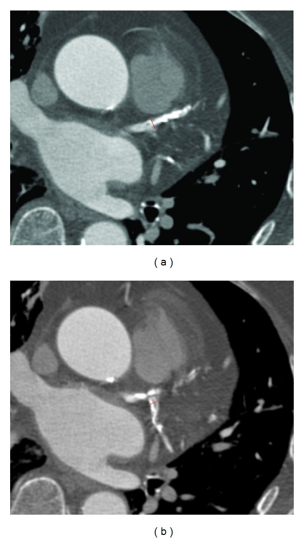
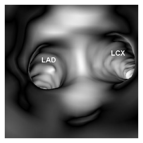
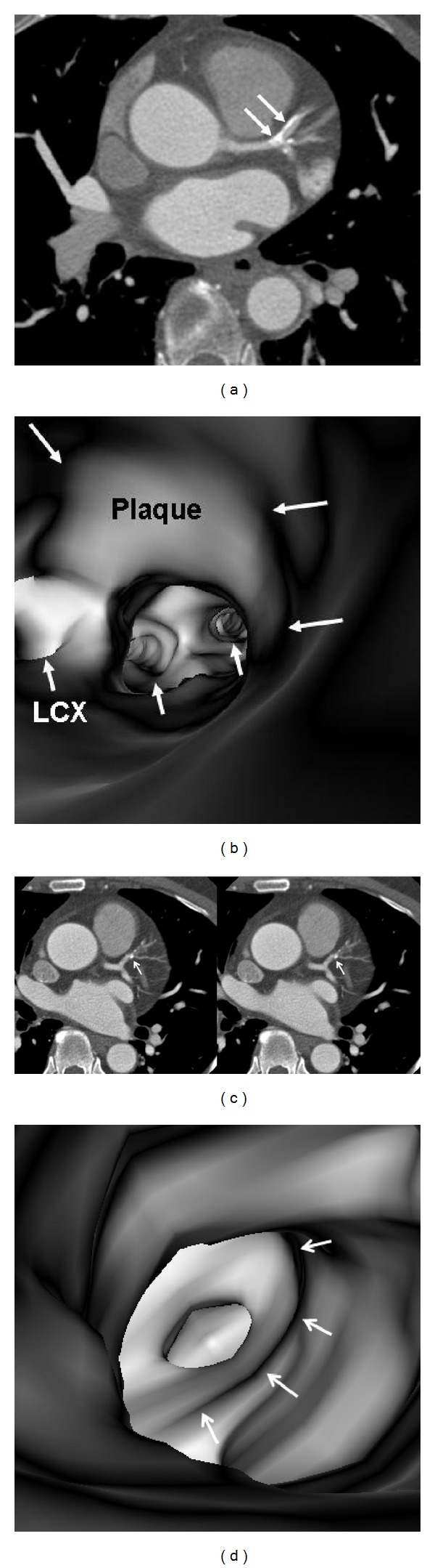
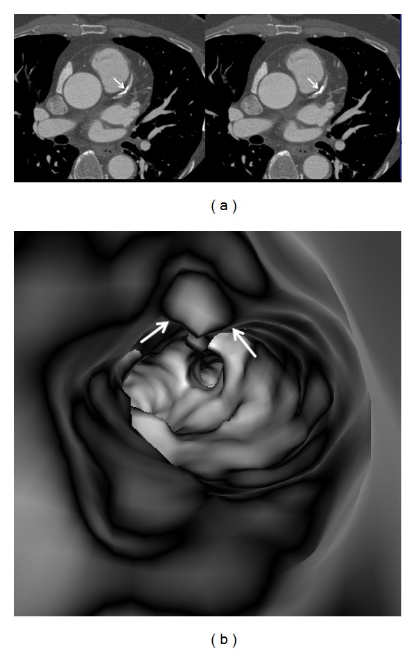
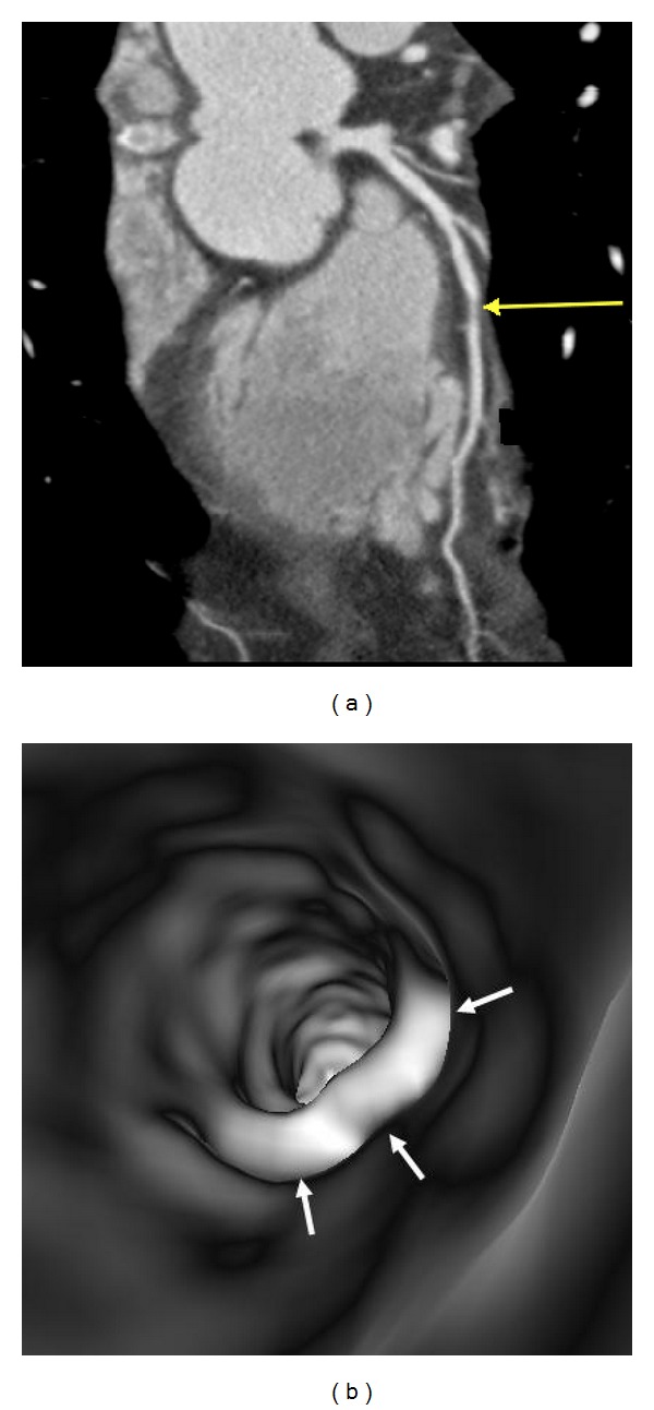
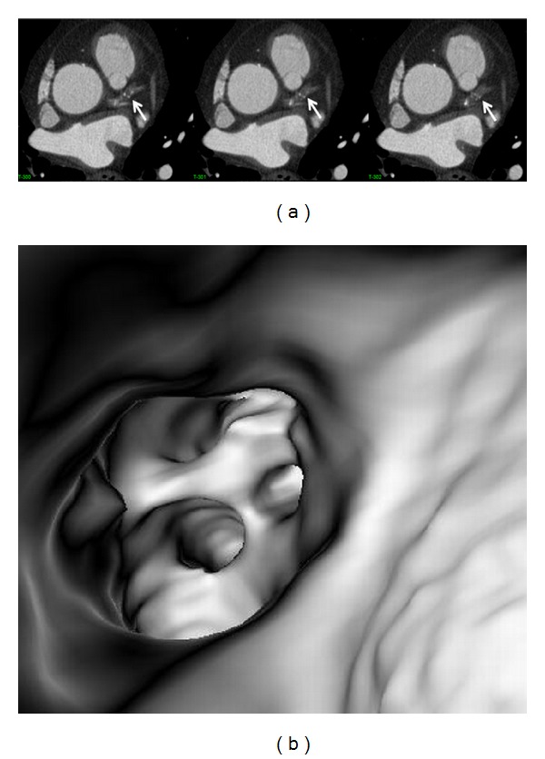
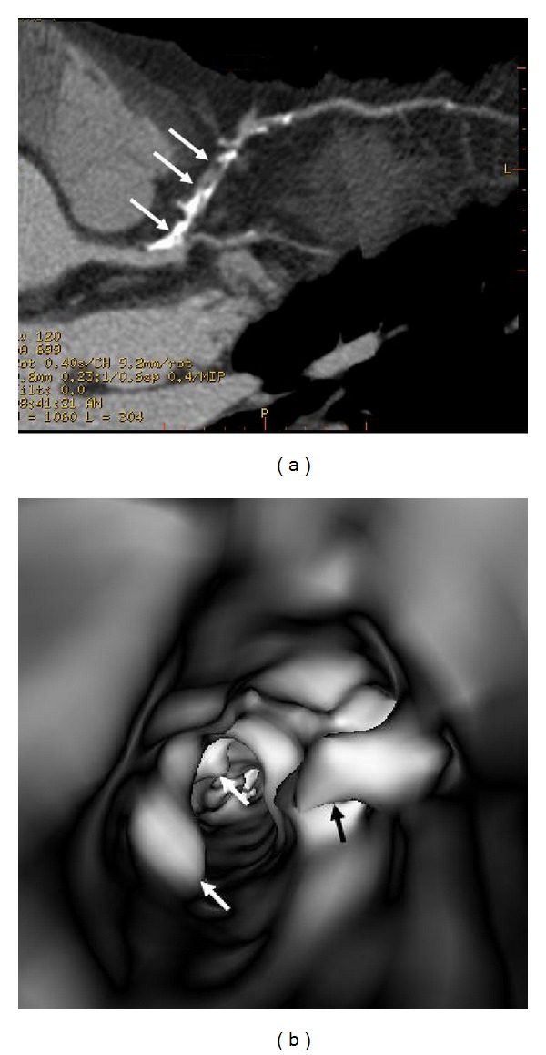
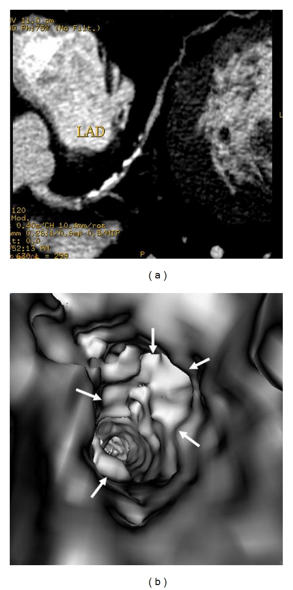
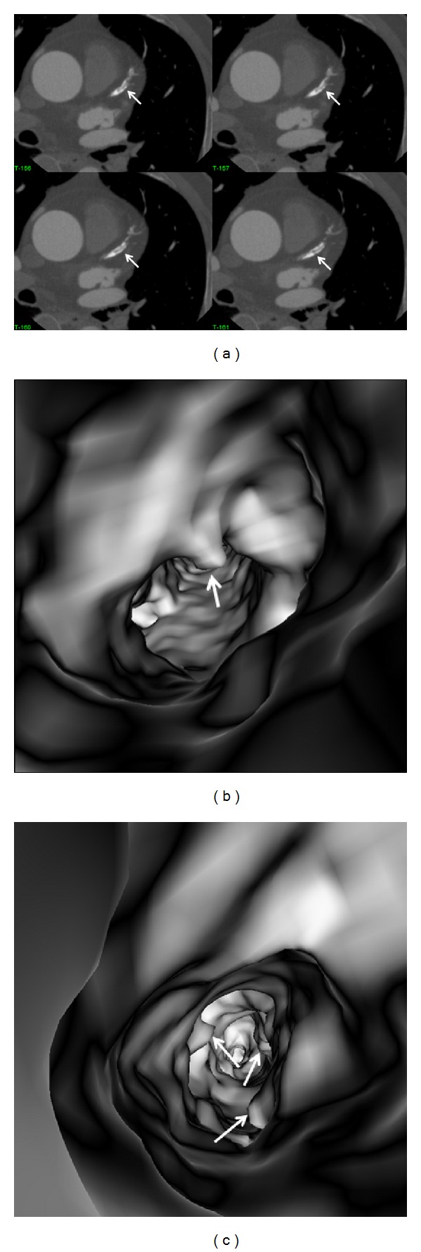
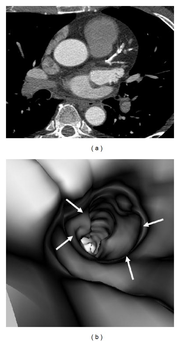
The aim of this study is to investigate the relationship between intraluminal appearances of coronary plaques and left coronary bifurcation angle and plaque components using coronary CT virtual intravascular endoscopy (VIE). Fifty patients suspected of coronary artery disease undergoing coronary CT angiography were included in the study. The left bifurcation angle in patients with diseased left coronary artery which was measured as 94.3° ± 16.5 is significantly larger than that in patients with normal left coronary artery, which was measured as 76.5° ± 15.9 (P < 0.001). Irregular VIE appearances were found in 10 out of 11 patients with mixed plaques in the left anterior descending (LAD) and left circumflex (LCx), while, in 29 patients with calcified plaques in the LAD and LCx, irregular VIE appearances were only noticed in 5 patients. Using 80° as a cut-off value to determine coronary artery disease, smooth VIE appearances were found in 95% of patients (18/19) with left bifurcation angle of less than 80°, while irregular VIE appearances were observed in nearly 50% of patients (15/31) with left bifurcation angle of more than 80°. This preliminary study shows that VIE appearances of the coronary lumen are directly related to the types of plaques.
Figures
References
-
- Raff GL, Gallagher MJ, O’Neill WW, Goldstein JA. Diagnostic accuracy of noninvasive coronary angiography using 64-slice spiral computed tomography. Journal of the American College of Cardiology. 2005;46(3):552–557. - PubMed
-
- Schuijf JD, Pundziute G, Jukema JW, et al. Diagnostic accuracy of 64-slice multislice computed tomography in the noninvasive evaluation of significant coronary artery disease. American Journal of Cardiology. 2006;98(2):145–148. - PubMed
-
- Rybicki FJ, Otero HJ, Steigner ML, et al. Initial evaluation of coronary images from 320-detector row computed tomography. International Journal of Cardiovascular Imaging. 2008;24(5):535–546. - PubMed
-
- Rodriguez-Granillo GA, Rosales MA, Degrossi E, Durbano I, Rodriguez AE. Multislice CT coronary angiography for the detection of burden, morphology and distribution of atherosclerotic plaques in the left main bifurcation. International Journal of Cardiovascular Imaging. 2007;23(3):389–392. - PubMed
-
- Mollet NR, Cademartiri F, van Mieghem CAG, et al. High-resolution spiral computed tomography coronary angiography in patients referred for diagnostic conventional coronary angiography. Circulation. 2005;112(15):2318–2323. - PubMed
MeSH terms
LinkOut - more resources
Full Text Sources
Other Literature Sources
Medical
