Review
. 2013:2013:193757.
doi: 10.1155/2013/193757. Epub 2013 Dec 17.

The role of autophagy in the pathogenesis of diabetic nephropathy

Affiliations
Review

The role of autophagy in the pathogenesis of diabetic nephropathy

Kosuke Yamahara et al. J Diabetes Res. 2013.

Abstract

Diabetic nephropathy is a leading cause of end-stage renal disease worldwide. The multipronged drug approach targeting blood pressure and serum levels of glucose, insulin, and lipids fails to fully prevent the onset and progression of diabetic nephropathy. Therefore, a new therapeutic target to combat diabetic nephropathy is required. Autophagy is a catabolic process that degrades damaged proteins and organelles in mammalian cells and plays a critical role in maintaining cellular homeostasis. The accumulation of proteins and organelles damaged by hyperglycemia and other diabetes-related metabolic changes is highly associated with the development of diabetic nephropathy. Recent studies have suggested that autophagy activity is altered in both podocytes and proximal tubular cells under diabetic conditions. Autophagy activity is regulated by both nutrient state and intracellular stresses. Under diabetic conditions, an altered nutritional state due to nutrient excess may interfere with the autophagic response stimulated by intracellular stresses, leading to exacerbation of organelle dysfunction and diabetic nephropathy. In this review, we discuss new findings showing the relationships between autophagy and diabetic nephropathy and suggest the therapeutic potential of autophagy in diabetic nephropathy.


Figures

Figure 1
Figure 1
Steps of the autophagic pathways. Five steps in the autophagic pathways have been identified: (1) initiation, isolated membrane appears in cytosol; (2) elongation, elongation is characterized by membrane bending and an increase in the size of the phagophore; (3) closure, the autophagosome membrane wraps around the cytosolic components; (4) fusion, the fusion of the autophagosome with a lysosome to form an autolysosome; and (5) breakdown, the autolysosome is degradated by lysosomal hydrolases.
Figure 2
Figure 2
Autophagy regulation. (a) ULK1 protein kinase complex. ULK1 is a critical regulator of nutrient-related autophagy. mTOR-dependent phosphorylation of ULK1 (Atg1) and Atg13 under nutrient-rich conditions inhibits autophagy. In contrast, AMPK-dependent phosphorylation of ULK1 activates autophagy induction under energy-depleted condition. (b) PI3K complex. Phosphatidylinositol 3-kinase (PI3K) phosphorylates phosphatidylinositol in the membrane lipid to create phosphatidylinositol 3-phosphate. Class III PI3K comprises hVps34, hVps15, Beclin-1, and Atg14. (c) Atg12-Atg5-Atg16 complex and LC3-II. Unlike other ubiquitin-like proteins, the ubiquitin-like protein Atg12 has a C-terminal glycine, which protects it from processing. Atg12 is conjugated to the substrate Atg5 by Atg7 and Atg10. The Atg12-Atg5 conjugate forms a complex with Atg16. Self-oligomerization of Atg16 results in a multimer of the Atg12-Atg5-Atg16 complex. After the ubiquitin-like protein LC3 has had its C-terminal arginine residue cleaved by the cysteine protease Atg4, it is passed on to Atg7 and Atg3 and transferred into the head group of its substrate phosphatidylethanolamine (PE). This LC3-PE conjugate functions as part of the membrane component of the autophagosome. When LC3-PE is once again deconjugated to PE by Atg4, Atg8 is recycled.
Figure 3
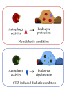
Figure 3
Autophagy in podocytes under diabetic conditions. Podocytes show high rates of autophagy even under nonstress conditions (upper panel). Autophagy activity is altered in podocytes under streptozotocin-induced type 1 diabetic conditions (lower panel). These results suggest that hyperglycemia alters autophagic activity, which may contribute to diabetes-related podocyte injury.
Figure 4
Figure 4
Autophagy in proximal tubular cells under diabetic conditions.   Proximal tubular cells show low rates of autophagy under basal conditions. Proteinuria renoprotectively elicits autophagy in proximal tubular cells (upper panel). Obesity suppresses proteinuria-induced autophagy via hyperactivation of mTORC1 in proximal tubular cells, leading to obesity-mediated exacerbation of proteinuria-induced tubulointerstitial damage (lower panel).

Similar articles

Cited by

References

    1. Burton C, Harris KPG. The role of proteinuria in the progression of chronic renal failure. American Journal of Kidney Diseases. 1996;27(6):765–775. - PubMed
    1. Abbate M, Zoja C, Remuzzi G. How does proteinuria cause progressive renal damage? Journal of the American Society of Nephrology. 2006;17(11):2974–2984. - PubMed
    1. Nath KA. Tubulointerstitial changes as a major determinant in the progression of renal damage. American Journal of Kidney Diseases. 1992;20(1):1–17. - PubMed
    1. Peterson JC, Adler S, Burkart JM, et al. Blood pressure control, proteinuria, and the progression of renal disease: the modification of diet in renal disease study. Annals of Internal Medicine. 1995;123(10):754–762. - PubMed
    1. Brenner BM, Cooper ME, De Zeeuw D, et al. Effects of losartan on renal and cardiovascular outcomes in patients with type 2 diabetes and nephropathy. The New England Journal of Medicine. 2001;345(12):861–869. - PubMed

Publication types