. 2013 Aug;1(2):123-33.
doi: 10.1158/2326-6066.CIR-13-0058.

Antigen-specific bacterial vaccine combined with anti-PD-L1 rescues dysfunctional endogenous T cells to reject long-established cancer

Affiliations

Antigen-specific bacterial vaccine combined with anti-PD-L1 rescues dysfunctional endogenous T cells to reject long-established cancer

David C Binder et al. Cancer Immunol Res. 2013 Aug.

Abstract

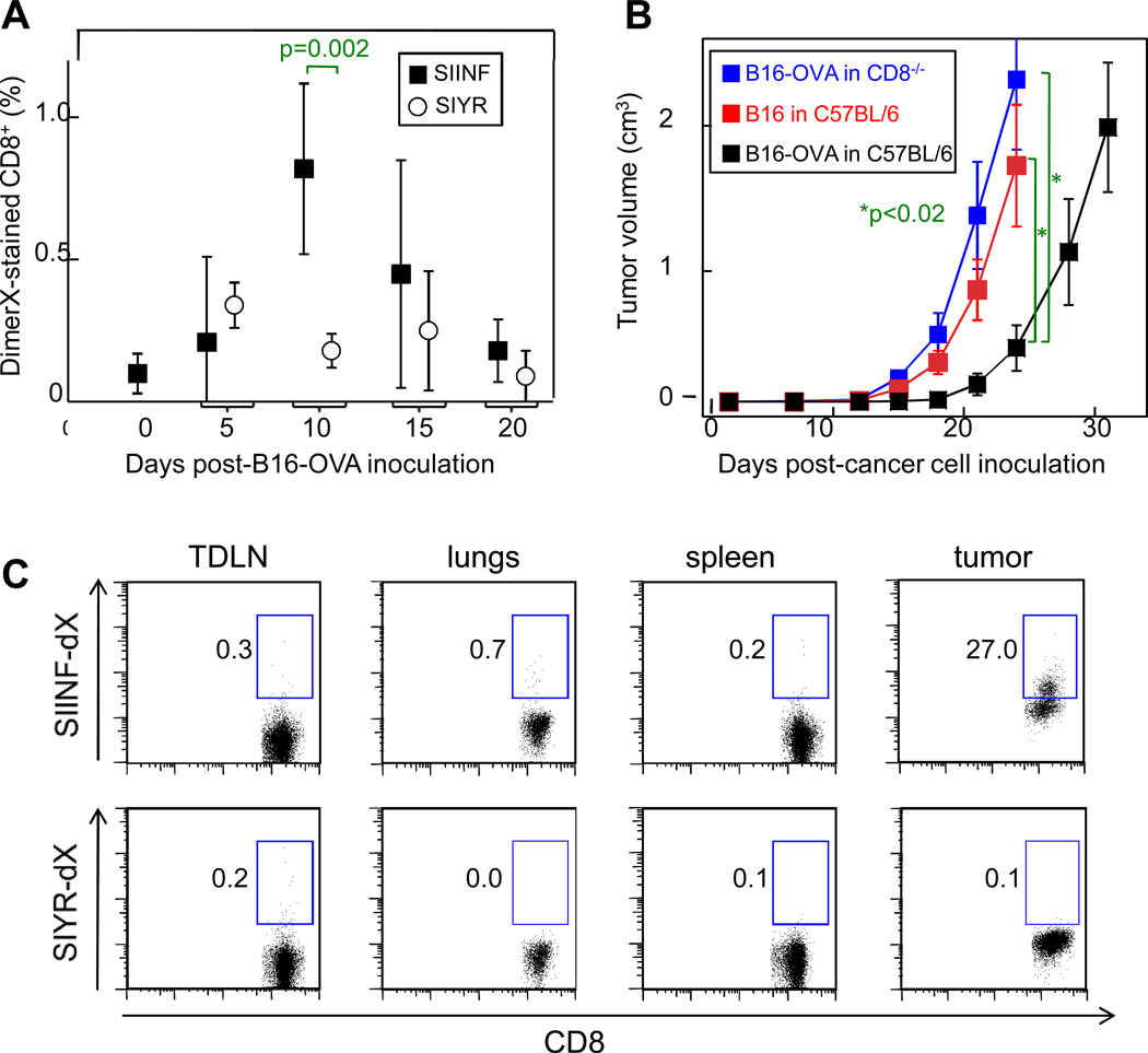
Immunogenic tumors grow progressively even when heavily infiltrated by CD8(+) T cells. We investigated how to rescue CD8(+) T cell function in long-established immunogenic melanomas that contained a high percentage of endogenous PD-1(+) tumor-specific CD8(+) T cells that were dysfunctional. Treatment with αPD-L1 and αCTLA-4 blocking antibodies did not prevent tumors from progressing rapidly. We then tested exogenous tumor-specific antigen delivery into tumors using Salmonella Typhimurium A1-R to increase antigen levels and generate a proinflammatory tumor microenvironment. Antigen-producing A1-R rescued the endogenous tumor-specific CD8(+) T cell response: proliferation was induced in the lymphoid organs and effector function was recovered in the tumor. Treatment with antigen-producing A1-R led to improved mouse survival and resulted in 32% rejection of long-established immunogenic melanomas. Following treatment with antigen-producing A1-R, the majority of tumor-specific CD8(+) T cells still expressed a high level of PD-1 in the tumor. Combining antigen-producing A1-R with αPD-L1 blocking antibody enhanced the expansion of tumor-specific CD8(+) T cells and resulted in 80% tumor rejection. Collectively, these data demonstrate a powerful new therapeutic approach to rescue dysfunctional endogenous tumor-specific CD8(+) T cells and eradicate advanced immunogenic tumors.

Keywords: CD8+ T cell rescue; PD-L1; S. Typhimurium; Tumor rejection; vaccine.


Conflict of interest statement

Ming Zhao and AntiCancer, Inc. hold patents for tumor-targeting bacteria. All other authors declare no conflict of interest.

Figures

Figure 1
Figure 1. A1-R SIINF delivers the SIINFEKL epitope into APCs for MHC Class I presentation
A) Diagram of the SIINF, SNFV, and EGFP constructs used in this study. B) High-copy number plasmids encoding the respective SIINF and SNFV fusion protein constructs were introduced into the A1-R strain. Whole bacterial lysates were examined for fusion protein expression using the anti-M45 antibody. C) J774 Kb-expressing macrophages were infected with A1-R SIINF or A1-R SNFV. Infected macrophages were then incubated with the B3Z (SIINFEKL-specific CD8+ T cell) hybridoma for 24 hours. B3Z stimulation was evaluated by the amount of IL-2 secreted into the culture as determined by ELISA. Data are representative of 2 independent experiments.
Figure 2
Figure 2. B16-OVA cancer cell inoculation induces priming of endogenous SIINF-specific CD8+ T cells that heavily infiltrate tumors
A) C57BL/6 mice were inoculated with B16-OVA cancer cells. At the indicated times, the peripheral blood leukocytes were stained with anti-CD8 and either SIINF/Kb-dimerX or control SIYR/Kb-dimerX. Data were pooled from 6 total mice with progressively growing tumors compiled from 2 independent experiments. B) B16-OVA and B16 tumor growth was measured following injection of 5 × 10⁶ cancer cells into C57BL/6 or C57BL/6 CD8−/− mice. The mean tumor volume (±SD) was calculated for each time-point. Each group consisted of 4 mice with progressively growing tumors; data are representative of 2 independent experiments. C) B16-OVA cancer cells were inoculated in a C57BL/6 mouse. 18 days later, the B16-OVA tumor reached 144 mm3. Single cell suspensions were made from the tumor-draining lymph node (TDLN), lungs, spleen, and tumor. The suspensions were stained with anti-CD8 and either SIINF/Kb-dimerX (SIINF-dX) or control SIYR/Kb-dimerX (SIYR-dX). Data are representative of 3 total mice from 2 independent experiments.
Figure 3
Figure 3. A1-R SIINF treatment rescues the SIINF-specific CD8+ T cell response in the blood and tumor
C57BL/6 mice bearing B16-OVA tumors were analyzed from the following groups: untreated tumors at day 0 (defined when tumors reached 100–168 mm3); untreated tumors at day 8; A1-R control-treated tumors 8–9 days post-treatment; and A1-R SIINF-treated tumors 8–9 days post-treatment. A) The peripheral blood was stained with anti-CD8 and SIINF/Kb-dimerX. The top panel is a representative stain from one mouse and the bottom panel contains pooled data with each mouse represented by a single dot. The A1-R control treatment group consists of 2 mice treated with A1-R SNFV and 2 mice treated with A1-R EGFP. ***p<0.001 when comparing A1-R SIINF to each other group. B) The upper two rows analyzed the percentage of SIINF-specific CD8+ T cells from tumors. Single cell suspensions from tumors were stained with anti-CD8 and either SIINF/Kb-dimerX or control SIYR/Kb-dimerX. The top panel is a representative stain from one mouse per group. The bottom panel contains pooled data from individual mice derived from at least 2 independent experiments per treatment group. The percentage of SIINF-specific CD8+ T cells represents the percentage of cells that stained positive with SIINF/Kb-dimerX subtracted by the background percentage of cells that stained positive with SIYR/Kb-dimerX. The difference between the A1-R SIINF group and each other group was non-significant (n.s.). The lower three rows analyzed cytokine production by SIINF-specific CD8+ T cells from the tumor. The same tumor cell suspensions, as analyzed in the upper two rows, were restimulated with SIINF peptide in the presence of Brefeldin A for 5 hrs. Cells were stained with SIINF/Kb-dimerX or control SIYR/Kb-dimerX, anti-CD8, anti-IFN-γ, and anti-TNF- α. Top panel shows a representative anti-IFN-γ and anti-TNF-α stain from gated SIINF/Kb-dimerX+ CD8+ double-positive cells. The bottom panels show pooled data. The percentage of IFN-γ+ or IFN-γ+TNF-α+ cells was defined as the percentage of SIINF-specific CD8+ T cells that stained positive with anti-IFN-γ and/or anti-TNF-α compared to the isotype controls. The A1-R control treatment group consisted of 4 mice treated with A1-R SNFV. ***p≤0.001 when comparing A1-R SIINF to each other group. **p≤0.002 when comparing A1-R OVA to each other group.
Figure 4
Figure 4. A1-R SIINF treatment results in a SIINF-specific CD8+ T cell-dependent antitumor effect on established B16-OVA tumors
C57BL/6 mice bearing established B16-OVA tumors were left untreated; treated one time with A1-R control; treated one time with A1-R SIINF; or treated one time with A1-R SIINF followed by treatment with the αCD8 depletion antibody. Lines indicate individual mice. The 4 mice that rejected the tumor following A1-R SIINF treatment were held for at least 100 days post-treatment. To determine if the tumors were eradicated, 3 of these mice were injected with 3 doses of 200 µg αCD8 at 3 day intervals. There was no tumor outgrowth for the 34 days following CD8+ T cell depletion before the mice were sacrificed. The A1-R control treatment group consisted of 3 mice treated with A1-R EGFP and 2 mice treated with A1-R SNFV. † indicates a single mouse that died during the experiment.
Figure 5
Figure 5. Treatment with A1-R SIINF combined with anti-PD-L1 results in enhanced SIINF-specific CD8+ T cell expansion and consistent tumor rejection
A) C57BL/6 mice bearing established B16-OVA tumors were treated as indicated. A blue dot represents the initial time of treatment with αCTLA-4 and αPD-L1 for an individual mouse. A red dot represents treatment with A1-R SIINF for an individual mouse. In each treatment group, mice were treated with 100 µg αCTLA-4 and/or 150 µg αPD-L1 every third day until the tumor relapsed completely (>1.5 cm3) or was rejected. Mice that rejected tumors were held for at least 100 days post-treatment prior to administration of 3 doses of 200 µg αCD8 at 3 day intervals. There was no tumor outgrowth for at least 40 days post-CD8+ T cell depletion before the mice were sacrificed, demonstrating that tumors were fully eradicated. B) At 8 or 9 days post-treatment, the peripheral blood from mice in the indicated treatment groups was stained with anti-CD8 and SIINF/Kb-dimerX. The percentage of SIINF-specific CD8+ T cells from mice that rejected B16-OVA versus relapsed following A1-R SIINF treatment was significantly different (p<0.01).

References

    1. Monach PA, Meredith SC, Siegel CT, Schreiber H. A unique tumor antigen produced by a single amino acid substitution. Immunity. 1995;2:45–59. - PubMed
    1. Coulie PG, Lehmann F, Lethe B, Herman J, Lurquin C, Andrawiss M, et al. A mutated intron sequence codes for an antigenic peptide recognized by cytolytic T lymphocytes on a human melanoma. Proc Natl Acad Sci U S A. 1995;92:7976–7980. - PMC - PubMed
    1. Wolfel T, Hauer M, Schneider J, Serrano M, Wolfel C, Klehmann-Hieb E, et al. A p16INK4a-insensitive CDK4 mutant targeted by cytolytic T lymphocytes in a human melanoma. Science. 1995;269:1281–1284. - PubMed
    1. Berendt MJ, North RJ. T-cell-mediated suppression of anti-tumor immunity. An explanation for progressive growth of an immunogenic tumor. J Exp Med. 1980;151:69–80. - PMC - PubMed
    1. Willimsky G, Blankenstein T. Sporadic immunogenic tumours avoid destruction by inducing T-cell tolerance. Nature. 2005;437:141–146. - PubMed

Publication types

MeSH terms