. 2014 Apr:310:1-12.
doi: 10.1016/j.heares.2014.01.003. Epub 2014 Jan 21.

A subset of chicken statoacoustic ganglion neurites are repelled by Slit1 and Slit2

Affiliations

A subset of chicken statoacoustic ganglion neurites are repelled by Slit1 and Slit2

Andrea C Battisti et al. Hear Res. 2014 Apr.

Abstract

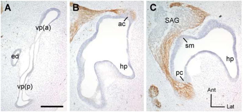
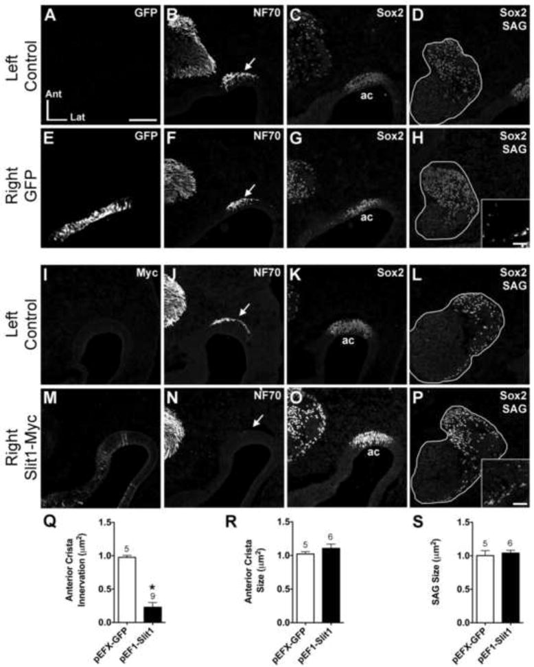
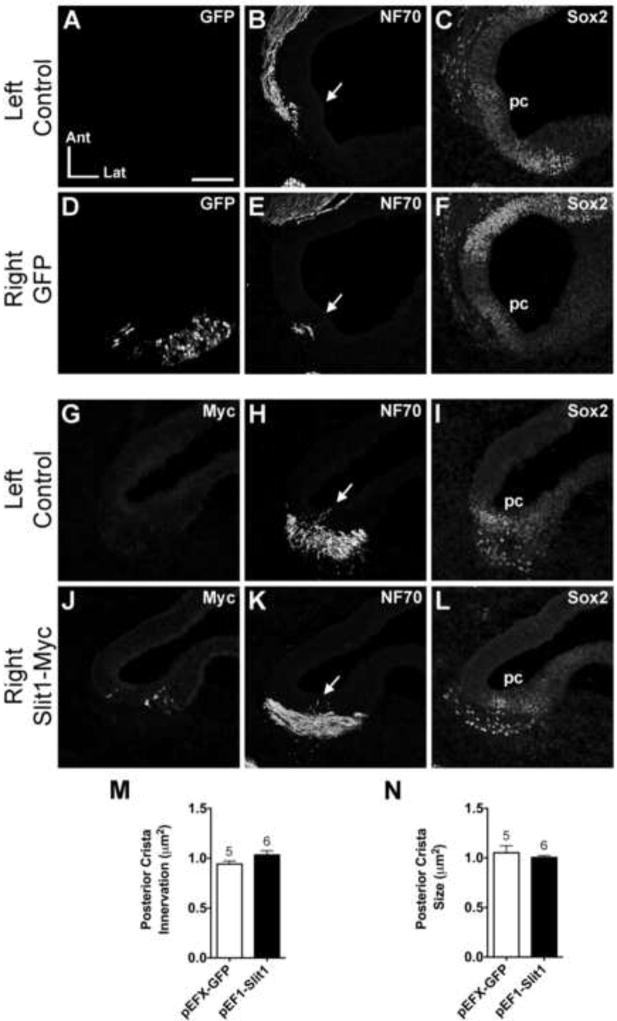
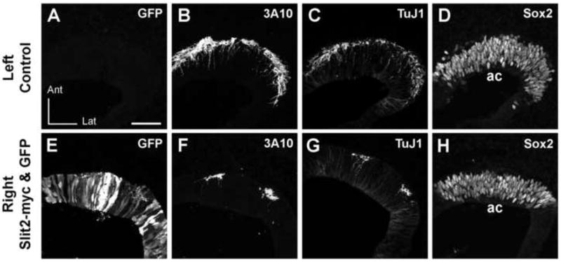
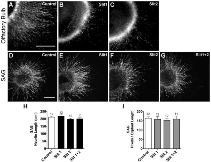
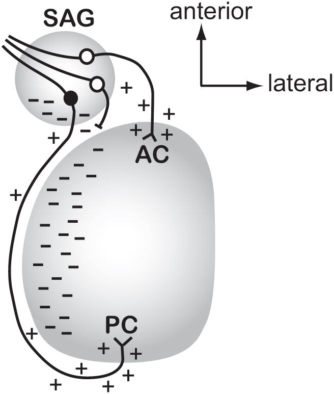
Mechanosensory hair cells in the chicken inner ear are innervated by bipolar afferent neurons of the statoacoustic ganglion (SAG). During development, individual SAG neurons project their peripheral process to only one of eight distinct sensory organs. These neuronal subtypes may respond differently to guidance cues as they explore the periphery in search of their target. Previous gene expression data suggested that Slit repellants might channel SAG neurites into the sensory primordia, based on the presence of robo transcripts in the neurons and the confinement of slit transcripts to the flanks of the prosensory domains. This led to the prediction that excess Slit proteins would impede the outgrowth of SAG neurites. As predicted, axonal projections to the primordium of the anterior crista were reduced 2-3 days after electroporation of either slit1 or slit2 expression plasmids into the anterior pole of the otocyst on embryonic day 3 (E3). The posterior crista afferents, which normally grow through and adjacent to slit expression domains as they are navigating towards the posterior pole of the otocyst, did not show Slit responsiveness when similarly challenged by ectopic delivery of slit to their targets. The sensitivity to ectopic Slits shown by the anterior crista afferents was more the exception than the rule: responsiveness to Slits was not observed when the entire E4 SAG was challenged with Slits for 40 h in vitro. The corona of neurites emanating from SAG explants was unaffected by the presence of purified human Slit1 and Slit2 in the culture medium. Reduced axon outgrowth from E8 olfactory bulbs cultured under similar conditions for 24 h confirmed bioactivity of purified human Slits on chicken neurons. In summary, differential sensitivity to Slit repellents may influence the directional outgrowth of otic axons toward either the anterior or posterior otocyst.


Figures

Figure 1
Figure 1. Horizontal histological sections show the plane of section used to evaluate canal morphogenesis and cristae innervation patterns
Frozen sections were immunostained with 3A10 antibody to detect the axons (brown) and counterstained with cresyl violet to visualize the otic epithelium. This is the untransfected (left) ear from an experimental embryo processed at HH28 3 days following electroporation into the contralateral ear. Images were reversed to match the orientation of specimens shown in Figures 2-4. This exemplar corresponds to the survival times of the embryo shown in figure 4. The embryos shown in Figures 2 and 3 were processed one day earlier, at 2-days post-electrorporation. These sections are arranged from dorsal to ventral (A-C). In panel (A), a morphologically normal vertical canal pouch is elongated along the anterior-posterior axis, flattened in the medial-lateral dimension as the walls of the pouch approach each other in advance of fusion, and has slight dilation of the rims of the pouch where the future canals will be. Panel B is a more ventral section, through the plane of axons projecting into the anterior crista primordium. Panel C is even more ventral to panel B. In this plane, some axons leave the statoacoustic ganglion (SAG) and immediately enter the primordium of the saccular macula. Others project well past this location to enter the posterior crista primordium. Abbreviations: ac, anterior crista; Ant, anterior; vp(a), vertical pouch, anterior part (will become the anterior semicircular canal); ed, endolymphatic duct; hp, horizontal pouch (will become the lateral semicircular canal); Lat, lateral; pc, posterior crista; vp(p), vertical pouch, posterior part (will become the posterior semicircular canal); SAG, statoacoustic ganglion; sm, saccular macula; pc, posterior crista. Scale bar = 200 microns.
Figure 2
Figure 2. Anterior crista afferents are repelled from the anterior crista following pEF1-hSlit1 electroporation
Serial horizontal sections through the left (unelectroporated) or right (electroporated) otocysts of embryos transfected with either pEFX-GFP (HH23; A-H) or pEF1-Slit1 (HH26; I-P). In these and subsequent figures, images of the left and right ear are from the same embryo, but the one side was mirror-image reversed to facilitate comparisons to the other side. Sections were immunostained with antibodies indicated in the upper right of each panel to label transfected cells (myc), axons (NF70) and prosensory domains (Sox2). Arrows indicate axon bundles adjacent to the anterior crista (ac) primordia. pEF1-Slit1 electroporation resulted in diffuse expression of Slit1-myc in the anterior crista primordium (M). The anterior crista of the Slit-electroporated ear (N) lacks afferent innervation as compared to the control ear of the same embryo (J). Sox2 marks the anterior crista primordium in each ear (C, G, K, O). The size and position of the SAGs (outlined in white) are similar across treatment groups (D, H, L, P). Insets in H and P show GFP- and Myc- immunolabeled cells in the SAGs, respectively. Quantification of anterior crista innervation (Q), anterior crista size (R), and SAG size (S). *p<0.0001 statistically significant from unelectroporated controls. The number above the bars represents the number of ears analyzed in each treatment group. Abbrevations: Ant, anterior; ac, anterior crista; Lat, lateral; SAG, statoacoustic ganglion. Scale bar in Panel A = 100 μm for all large panels. Scale bar for insets in Panel H = 25 μm.
Figure 3
Figure 3. Normal posterior crista innervation following pEF1-Slit1 electroporation
Serial horizontal sections through the left and right posterior cristae of pEFX-GFP (HH24; A-F) and pEF1-Slit1 (HH26; G-L) electroporated embryos. Arrows indicate projections into the posterior crista primordium. Innervation of the Slit1-electroporated posterior crista (K) resembles the control ear (H). Sox2 labels the posterior crista in each ear (C, F, I, L). Quantification of posterior crista innervation (M) and size (N). See legend of Figure 2 for labeling conventions. Abbreviations: Ant, anterior; Lat, lateral; pc, posterior crista; SAG, statoacoustic ganglion. Scale bar = 100 μm.
Figure 4
Figure 4. Anterior crista afferents are reduced 3 days after co-electroporation of pEF1-Slit2 and pEFX-GFP in their target domain
Serial horizontal sections through the left or right otocysts of an embryo in which both pEFX-GFP and pEF1-Slit2 were co-electroporated into the right ear. The same sections are shown in alternate color channels for panels A/B and C/D, while the remaining panels are from sequential sections through each ear. Images of the right ear were mirror-image reversed to facilitate comparing to the left ear. The antibodies used are indicated in the upper right of each panel. GFP is undetectable in the left ear (A) but is present in a subset of cells distributed throughout the anterior pole of the right ear (E). Robust innervation of the anterior crista primordium of the left ear (B, C) can be contrasted with the paucity of axons detected beneath and within the anterior crista of the right ear (F,G). In the right ear, two axon bundles flank an area of particularly strong GFP expression in the epithelium that is completely devoid of axonal projections. Sox2 labeling reveals the anterior crista primordium on each side (D,H). Abbrevations: ac, anterior crista; Ant, anterior; Lat, lateral. Scale bar = 50 μm.
Figure 5
Figure 5. Purified Slit1 and Slit2 proteins inhibit HH33-34 chick olfactory bulb neurite outgrowth but do not affect HH20-25 SAG neurite outgrowth
Olfactory bulb explants cultured for 24 hours with either 20 μg/ml mouse Slit1 (B) or 20 μg/ml mouse Slit2 (C) display reduced neurite outgrowth compared to explants cultured without Slit (Control; A), confirming that the mouse Slit proteins are bioactive under these culture conditions. SAG explants display similar neurite outgrowth patterns when cultured for 40 hours without (D) or with Slit1 (E), Slit2 (F) or Slit1+2 (G). Quantification of SAG neurite length (H) and pixel number (I). Bars represent the mean for each treatment group (±SE). The number of explants analyzed, for each treatment group, is above the bars. Pixel data partially overlap with samples used for length measurements. Scale bars = 200 μm.
Figure 6
Figure 6. Model for how Slit activity may direct afferent outgrowth into the otocyst in vivo
This figure predicts where Slit1 and/or Slit2 proteins (indicated as dashes) are localized in the normal ear at the time when afferents to the anterior and posterior crista are projecting from the SAG towards their targets (approximately HH17-22). This model takes into account the experimental observation that afferents projecting to the anterior vs. posterior cristae demonstrate differential responsiveness to Slits as a mechanism by which these two populations of axons are directed to grow in opposite directions. We predict that the diffusion of Slit proteins (indicated by dashes) from the posterior SAG and medial otic vesicle will repel anterior crista afferents (white cell bodies) from initiating trajectories in the posterior direction. If they do project posteriorly, they would be prevented from advancing (shown as an axon with a blocked process) and may then be redirected anteriorly to reach their target. In contrast, a neuron seeking to innervate the posterior crista (black cell body) is insensitive to the same Slit cues, allowing it to pass through or alongside territories that have an abundance of Slit proteins. The model also depicts putative attractive cues (shown as + symbols) along the pathways or emanating from the targets, although additional unknown repulsive cues (not shown) may also confine axonal trajectories to specific pathways. Abbreviations: AC, anterior crista; PC, posterior crista; SAG, statoacoustic ganglion.

References

    1. Adamska M, Herbrand H, Adamski M, Kruger M, Braun T, Bober E. FGFs control the patterning of the inner ear but are not able to induce the full ear program. Mech Dev. 2001;109:303–313. - PubMed
    1. Agarwala S, Sanders TA, Ragsdale CW. Sonic hedgehog control of size and shape in midbrain pattern formation. Science. 2001;291:2147–2150. - PubMed
    1. Appler JM, Goodrich LV. Connecting the ear to the brain: Molecular mechanisms of auditory circuit assembly. Prog Neurobiol. 2011;93:488–508. - PMC - PubMed
    1. Bank LM, Bianchi LM, Ebisu F, Lerman-Sinkoff D, Smiley EC, Shen YC, Ramamurthy P, Thompson DL, Roth TM, Beck CR, et al. Macrophage migration inhibitory factor acts as a neurotrophin in the developing inner ear. Development. 2012;139:4666–74. - PMC - PubMed
    1. Bashaw GJ, Goodman CS. Chimeric axon guidance receptors: the cytoplasmic domains of slit and netrin receptors specify attraction versus repulsion. Cell. 1999;97:917–926. - PubMed

Publication types

MeSH terms