

Transplantation of umbilical cord-derived mesenchymal stem cells into the striata of R6/2 mice: behavioral and neuropathological analysis
- PMID: 24456799
- PMCID: PMC3854759
- DOI: 10.1186/scrt341
Transplantation of umbilical cord-derived mesenchymal stem cells into the striata of R6/2 mice: behavioral and neuropathological analysis
Abstract
Introduction: Huntington's disease (HD) is an autosomal dominant disorder caused by an expanded CAG repeat on the short arm of chromosome 4 resulting in cognitive decline, motor dysfunction, and death, typically occurring 15 to 20 years after the onset of motor symptoms. Neuropathologically, HD is characterized by a specific loss of medium spiny neurons in the caudate and the putamen, as well as subsequent neuronal loss in the cerebral cortex. The transgenic R6/2 mouse model of HD carries the N-terminal fragment of the human HD gene (145 to 155 repeats) and rapidly develops some of the behavioral characteristics that are analogous to the human form of the disease. Mesenchymal stem cells (MSCs) have shown the ability to slow the onset of behavioral and neuropathological deficits following intrastriatal transplantation in rodent models of HD. Use of MSCs derived from umbilical cord (UC) offers an attractive strategy for transplantation as these cells are isolated from a noncontroversial and inexhaustible source and can be harvested at a low cost. Because UC MSCs represent an intermediate link between adult and embryonic tissue, they may hold more pluripotent properties than adult stem cells derived from other sources.
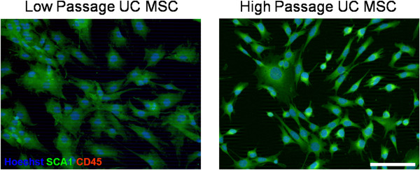
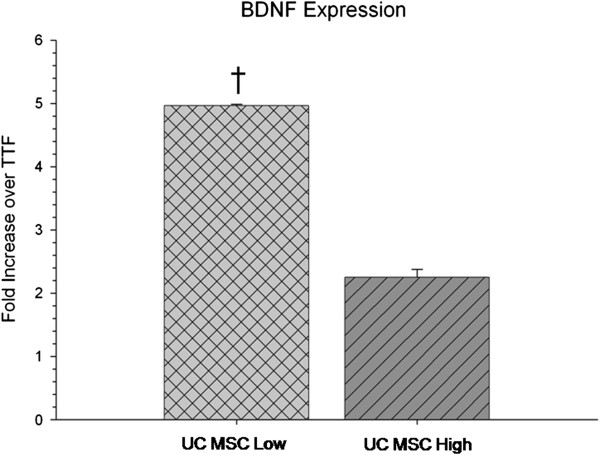
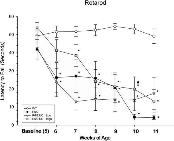
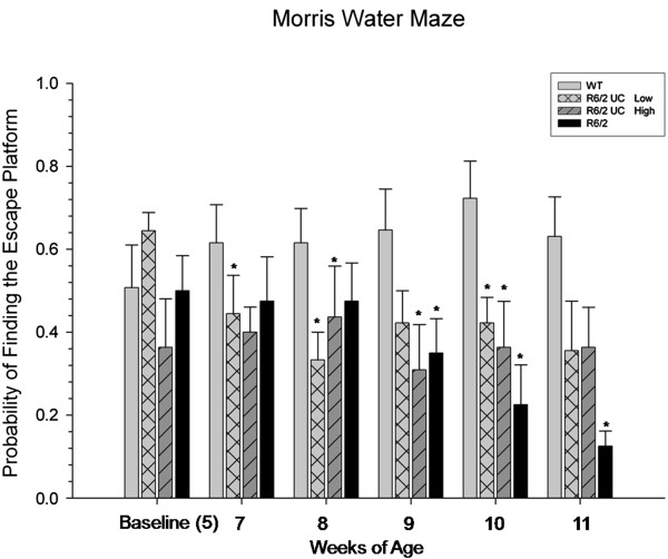
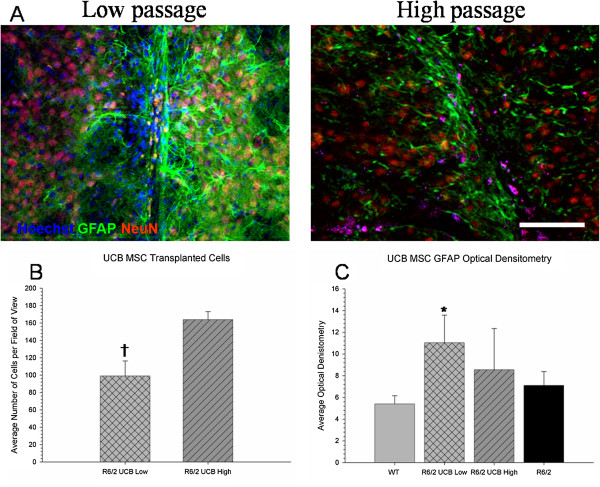
Methods: Mesenchymal stem cells, isolated from the UC of day 15 gestation pups, were transplanted intrastriatally into 5-week-old R6/2 mice at either a low-passage (3 to 8) or high-passage (40 to 50). Mice were tested behaviorally for 6 weeks using the rotarod task, the Morris water maze, and the limb-clasping response. Following behavioral testing, tissue sections were analyzed for UC MSC survival, the immune response to the transplanted cells, and neuropathological changes.
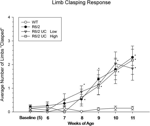
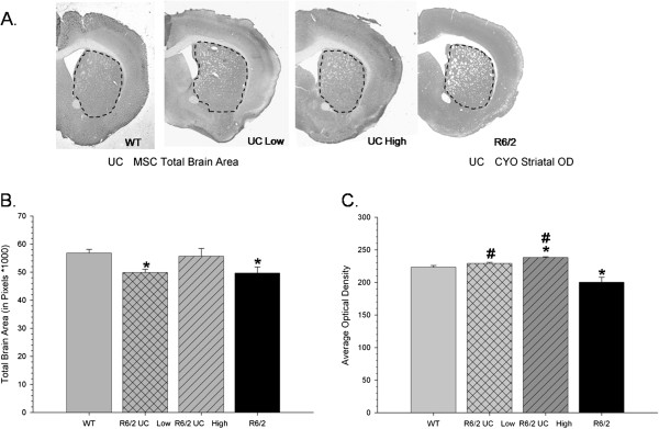
Results: Following transplantation of UC MSCs, R6/2 mice did not display a reduction in motor deficits but there appeared to be transient sparing in a spatial memory task when compared to untreated R6/2 mice. However, R6/2 mice receiving either low- or high-passage UC MSCs displayed significantly less neuropathological deficits, relative to untreated R6/2 mice.
Conclusions: The results from this study demonstrate that UC MSCs hold promise for reducing the neuropathological deficits observed in the R6/2 rodent model of HD.
Figures
Similar articles
-
Reductions in behavioral deficits and neuropathology in the R6/2 mouse model of Huntington's disease following transplantation of bone-marrow-derived mesenchymal stem cells is dependent on passage number.Stem Cell Res Ther. 2015 Feb 19;6(1):9. doi: 10.1186/scrt545. Stem Cell Res Ther. 2015. PMID: 25971780 Free PMC article.
-
Intranasal Administration of Mesenchymal Stem Cells Ameliorates the Abnormal Dopamine Transmission System and Inflammatory Reaction in the R6/2 Mouse Model of Huntington Disease.Cells. 2019 Jun 15;8(6):595. doi: 10.3390/cells8060595. Cells. 2019. PMID: 31208073 Free PMC article.
-
Genetically engineered mesenchymal stem cells reduce behavioral deficits in the YAC 128 mouse model of Huntington's disease.Behav Brain Res. 2010 Dec 25;214(2):193-200. doi: 10.1016/j.bbr.2010.05.023. Epub 2010 May 21. Behav Brain Res. 2010. PMID: 20493905
-
Umbilical cord-derived mesenchymal stem cells in neurodegenerative disorders: from literature to clinical practice.Regen Med. 2020 Apr;15(4):1561-1578. doi: 10.2217/rme-2019-0119. Epub 2020 Jun 1. Regen Med. 2020. PMID: 32479211 Review.
-
The application of umbilical cord-derived MSCs in cardiovascular diseases.J Cell Mol Med. 2021 Sep;25(17):8103-8114. doi: 10.1111/jcmm.16830. Epub 2021 Aug 11. J Cell Mol Med. 2021. PMID: 34378345 Free PMC article. Review.
Cited by
-
A novel method to isolate mesenchymal stem cells from mouse umbilical cord.Mol Med Rep. 2018 Jan;17(1):861-869. doi: 10.3892/mmr.2017.7950. Epub 2017 Nov 3. Mol Med Rep. 2018. PMID: 29115623 Free PMC article.
-
Mesenchymal Stem Cells for Neurological Disorders.Adv Sci (Weinh). 2021 Feb 24;8(7):2002944. doi: 10.1002/advs.202002944. eCollection 2021 Apr. Adv Sci (Weinh). 2021. PMID: 33854883 Free PMC article. Review.
-
Prion-like mechanisms in neurodegenerative disease: Implications for Huntington's disease therapy.Stem Cells Transl Med. 2020 May;9(5):559-566. doi: 10.1002/sctm.19-0248. Epub 2020 Jan 30. Stem Cells Transl Med. 2020. PMID: 31997581 Free PMC article. Review.
-
Progress in developing transgenic monkey model for Huntington's disease.J Neural Transm (Vienna). 2018 Mar;125(3):401-417. doi: 10.1007/s00702-017-1803-y. Epub 2017 Nov 10. J Neural Transm (Vienna). 2018. PMID: 29127484 Free PMC article. Review.
-
Advancements in Umbilical Cord Biobanking: A Comprehensive Review of Current Trends and Future Prospects.Stem Cells Cloning. 2024 Dec 5;17:41-58. doi: 10.2147/SCCAA.S481072. eCollection 2024. Stem Cells Cloning. 2024. PMID: 39655226 Free PMC article. Review.
References
-
- Ende N, Chen R. Human umbilical cord blood cells ameliorate Huntington’s disease in transgenic mice. J Med. 2001;32:231–240. - PubMed
Publication types
MeSH terms
Substances
LinkOut - more resources
Full Text Sources
Other Literature Sources
Medical
