. 2014 Jan 24:11:5.
doi: 10.1186/1743-8977-11-5.

Equivalent titanium dioxide nanoparticle deposition by intratracheal instillation and whole body inhalation: the effect of dose rate on acute respiratory tract inflammation

Affiliations

Equivalent titanium dioxide nanoparticle deposition by intratracheal instillation and whole body inhalation: the effect of dose rate on acute respiratory tract inflammation

Brittany L Baisch et al. Part Fibre Toxicol. .

Abstract

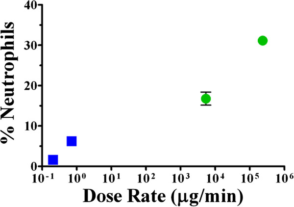
Background: The increased production of nanomaterials has caused a corresponding increase in concern about human exposures in consumer and occupational settings. Studies in rodents have evaluated dose-response relationships following respiratory tract (RT) delivery of nanoparticles (NPs) in order to identify potential hazards. However, these studies often use bolus methods that deliver NPs at high dose rates that do not reflect real world exposures and do not measure the actual deposited dose of NPs. We hypothesize that the delivered dose rate is a key determinant of the inflammatory response in the RT when the deposited dose is constant.

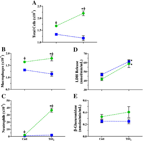
Methods: F-344 rats were exposed to the same deposited doses of titanium dioxide (TiO₂) NPs by single or repeated high dose rate intratracheal instillation or low dose rate whole body aerosol inhalation. Controls were exposed to saline or filtered air. Bronchoalveolar lavage fluid (BALF) neutrophils, biochemical parameters and inflammatory mediator release were quantified 4, 8, and 24 hr and 7 days after exposure.

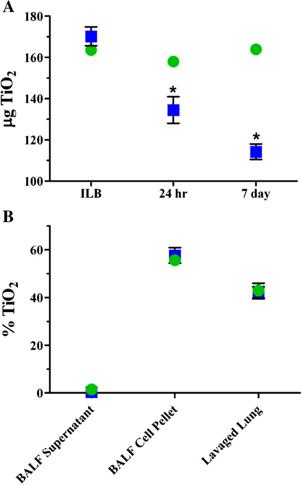
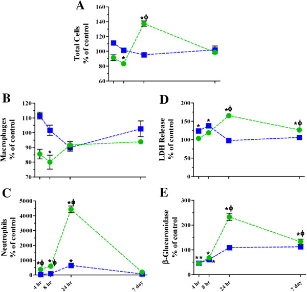
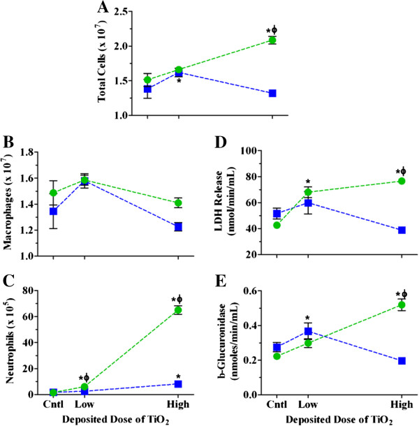
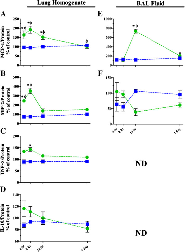
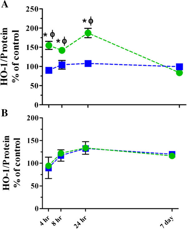
Results: Although the initial lung burdens of TiO₂ were the same between the two methods, instillation resulted in greater short term retention than inhalation. There was a statistically significant increase in BALF neutrophils at 4, 8 and 24 hr after the single high dose TiO₂ instillation compared to saline controls and to TiO₂ inhalation, whereas TiO₂ inhalation resulted in a modest, yet significant, increase in BALF neutrophils 24 hr after exposure. The acute inflammatory response following instillation was driven primarily by monocyte chemoattractant protein-1 and macrophage inflammatory protein-2, mainly within the lung. Increases in heme oxygenase-1 in the lung were also higher following instillation than inhalation. TiO₂ inhalation resulted in few time dependent changes in the inflammatory mediator release. The single low dose and repeated exposure scenarios had similar BALF cellular and mediator response trends, although the responses for single exposures were more robust.

Conclusions: High dose rate NP delivery elicits significantly greater inflammation compared to low dose rate delivery. Although high dose rate methods can be used for quantitative ranking of NP hazards, these data caution against their use for quantitative risk assessment.


Figures

Figure 1
Figure 1
Dosimetry and short term retention of TiO2 for single high dose TiO2 exposure. The mass of TiO2 deposited in the lung following whole body inhalation (blue squares) and intratracheal instillation (green circles) was determined immediately, 24 hr and 7 days post exposure (A) and the compartmentalization of TiO2 in BALF supernatant, BALF cell pellet and the remaining lung tissue after lavage (lavaged lung) were determined 24 hr post exposure (B). Values are group means (n = 3–5) ± standard error (SE). The inhalation and instillation ILBs were not found to be significantly different; *, significantly different compared to the respective ILB (p < 0.05).
Figure 2
Figure 2
Time course of changes in BALF cellular and biochemical parameters following single high dose exposure to TiO2 NPs. The numbers of cells (A), macrophages (B), and neutrophils (C), and LDH (D) and β-glucuronidase activities (E) were assessed 4, 8, 24 hr and 7 days post inhalation (blue squares) or instillation (green circles) exposure. Values are group means ± SE (n = 5) and are represented as a percentage of respective controls. *, significantly different from corresponding controls; Φ, significantly different between exposure methods (p < 0.05).
Figure 3
Figure 3
Dose response relationships for BALF cellular and biochemical parameters 24 hr post exposure to TiO2 NPs. The numbers of cells (A), macrophages (B), and neutrophils (C), and LDH (D) and β-glucuronidase activities (E) 24 hr post inhalation (blue squares) or instillation (green circles) exposure to increasing deposited doses of TiO2 NPs. Values are group means ± SE (n = 5) and are represented as a percentage of respective controls. *, significantly different from corresponding controls; Φ, significantly different between exposure methods (p < 0.05).
Figure 4
Figure 4
Inflammatory mediator release in lung homogenates and BALF supernatants following single high dose exposure to TiO2 NPs. Changes in MCP-1 (A, homogenates;E, BALF supernatants), MIP-2 (B, homogenates;F, BALF supernatants), TNF-α (C, homogenates) and IL-10, (D, homogenates) following inhalation (blue squares) or instillation (green circles) are graphed as percentage of controls. Values are group means ± SE (n = 5). ND, levels were below the limit of detection by ELISA (TNF-α and IL-10 ~15 pg/mL). *, significantly different from corresponding controls; Φ, significantly different between exposure methods (p < 0.05).
Figure 5
Figure 5
HO-1 levels in lung homogenates and BALF cell pellets following single high dose exposure to TiO2 NPs. HO-1 levels in lung homogenates (A) and BALF cell pellets (B) following inhalation (blue squares) or instillation (green circles) exposure. Values are group means ± SE (n = 5) and represented as percentage of controls. *, significantly different from corresponding controls; Φ, significantly different between exposure methods (p < 0.05).
Figure 6
Figure 6
BALF cellular and biochemical parameters following repeated high dose exposure to TiO2 NPs. The numbers of cells (A), macrophages (B), and neutrophils (C), and LDH (D) and β-glucuronidase activities (E) were assessed 24 hr post inhalation (blue squares) or instillation (green circles) exposure. Values are group means ± SE (n = 5). *, significantly different from corresponding controls; Φ, significantly different between exposure methods (p < 0.05).
Figure 7
Figure 7
Percentage of neutrophils 24 hr after single and repeated exposure to high dose TiO2 at different dose rates. The percentage of neutrophils in BALF at 24 hr post inhalation (blue squares) or instillation (green circles) exposure plotted according to deposited dose rate (~0.21, repeated inhalation; ~0.71, high dose single inhalation; ~5,400, repeated instillation; and ~240,000 μg/min, high dose single instillation). Symbols are group means ± SE (n = 5).

Similar articles

Cited by

References

    1. Nel A, Xia T, Madler L, Li N. Toxic potential of materials at the nanolevel. Science. 2006;311:622–627. doi: 10.1126/science.1114397. - DOI - PubMed
    1. Oberdörster G, Stone V, Donaldson K. Toxicology of nanoparticles: a historical perspective. Nanotoxicology. 2007;1:2–25. doi: 10.1080/17435390701314761. - DOI
    1. Project on emerging nanotechnologies. http:/www.nanotechproject.org/cpi.
    1. Rushton EK, Jiang J, Leonard SS, Eberly S, Castranova V, Biswas P, Elder A, Han X, Gelein R, Finkelstein J, Oberdörster G. Concept of assessing nanoparticle hazards considering nanoparticle dosemetric and chemical/biological response metrics. J Toxicol Environ Health A. 2010;73:445–461. doi: 10.1080/15287390903489422. - DOI - PMC - PubMed
    1. Xia T, Hamilton RF, Bonner JC, Crandall ED, Elder A, Fazlollahi F, Girtsman TA, Kim K, Mitra S, Ntim SA. et al.Interlaboratory evaluation of in vitro cytotoxicity and inflammatory responses to engineered nanomaterials: the NIEHS Nano GO Consortium. Environ Health Perspect. 2013;121:683–690. doi: 10.1289/ehp.1306561. - DOI - PMC - PubMed

Publication types

MeSH terms