. 2014 Feb;24(2):97-116.
doi: 10.1016/j.nmd.2013.11.003. Epub 2013 Nov 18.

Approach to the diagnosis of congenital myopathies

Collaborators, Affiliations

Approach to the diagnosis of congenital myopathies

Kathryn N North et al. Neuromuscul Disord. 2014 Feb.

Abstract

Over the past decade there have been major advances in defining the genetic basis of the majority of congenital myopathy subtypes. However the relationship between each congenital myopathy, defined on histological grounds, and the genetic cause is complex. Many of the congenital myopathies are due to mutations in more than one gene, and mutations in the same gene can cause different muscle pathologies. The International Standard of Care Committee for Congenital Myopathies performed a literature review and consulted a group of experts in the field to develop a summary of (1) the key features common to all forms of congenital myopathy and (2) the specific features that help to discriminate between the different genetic subtypes. The consensus statement was refined by two rounds of on-line survey, and a three-day workshop. This consensus statement provides guidelines to the physician assessing the infant or child with hypotonia and weakness. We summarise the clinical features that are most suggestive of a congenital myopathy, the major differential diagnoses and the features on clinical examination, investigations, muscle pathology and muscle imaging that are suggestive of a specific genetic diagnosis to assist in prioritisation of genetic testing of known genes. As next generation sequencing becomes increasingly used as a diagnostic tool in clinical practise, these guidelines will assist in determining which sequence variations are likely to be pathogenic.

Keywords: Congenital myopathy; Diagnosis; Guidelines.


Conflict of interest statement

We declare that the authors have no conflict of interest or financial relationship with this work.

Figures

Fig. 1
Fig. 1
Facial involvement in congenital myopathies. (A) Pronounced facial weakness, particularly affecting the lower face and mouth resulting in craniofacial dysmorphism (“myopathic facies”) in sisters aged 6 years and 3 months with autosomal recessive nemaline myopathy (likely due to nebulin). (B) Ptosis and ophthalmoplegia in a patient with DNM2-related centronuclear myopathy at age 9 years.
Fig. 2
Fig. 2
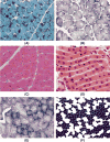
Pathological features that define the major subtypes of congenital myopathy. (A) Nemaline rods: Biopsy from patient with nemaline myopathy with a dominant mutation in the ACTA1 gene, showing clusters of purple staining rods at the periphery of most fibres and some internal within fibres (Gomori trichrome). (B) Central cores: Biopsy of the quadriceps from a three year old patient with central core disease with a dominant mutation in the ryanodine receptor gene showing mild variation in fibre size (fibre diameter range 15–65 mm), fibre type uniformity and numerous cores of varying size centrally or peripherally (oxidative enzyme stain SDH). (C) Central nuclei: Quadriceps biopsy from a 28 year old patient with autosomal dominant centronuclear myopathy due to a DNM2 mutation. The biopsy demonstrated small type 1 fibres and centrally placed nuclei in the majority of fibres (H&E). (D) Central nuclei (longitudinal section): Quadriceps biopsy from a case of X-linked myotubular myopathy aged 8 months showing large central nuclei. Note the widely spaced nuclei which affects the number seen in transverse section Most fibres are less than 10 mm in diameter. (E) Multiminicores: Areas in both fibre types of varying size and number devoid of oxidative enzyme stain in a quadriceps biopsy from a case of ‘multi-minicore disease’ aged 11 years with recessive mutations in the SEPN1 gene (NADH-TR). (F) Congenital fibre type disproportion with mutation in ACTA1: The only apparent pathology in this case was the small size of the dark-staining type 1 fibres and type 1 fibre predominance (ATPase preincubated at pH 4.3). Fibre diameter 25–70 mm.
Fig. 3
Fig. 3
Muscle magnetic resonance imaging (MRI) in the diagnosis of the congenital myopathies: T1-weighted images, transverse sections, through the thigh (A–D) and the lower leg (E–H) in a normal control (A,E) and patients with RYR1-related dominant central core disease (CCD) (B,F), DNM2-related dominant centronuclear myopathy (CNM) (C,G) and NEB-related recessive nemaline myopathy (NM) (D,H). In RYR1-related dominant CCD (B,F), there is marked involvement of the thigh with relative sparing of rectus femoris (RF), adductor longus (AL), gracilis (G) and hamstring muscles. In the lower leg, soleus (So), gastrocnemius lateralis (Gl) and peroneal group (PG) are markedly affected whereas tibialis anterior (AT), tibialis posterior (TP) and gastrocnemius medialis (Gm) are relatively spared. In DNM2-related dominant centronuclear myopathy (CNM) (C,G), the thigh is diffusely affected with prominent involvement of rectus femoris (RF) and adductor longus (AL) compared to other muscle groups. In the lower leg, there is diffuse involvement of all muscle groups with relative sparing of the tibialis posterior (TP). In NEB-related recessive nemaline myopathy (NM) (D,H), the thigh is spared whereas the lower leg shows early involvement of the tibialis anterior (AT) and, to a lesser extent, soleus (So), in keeping with the clinical finding of early distal involvement in NM. (Composite image from [74,85,93]).

References

    1. Laing NG, Wallgren-Pettersson C. 161st ENMC International Workshop on nemaline myopathy and related disorders, Newcastle upon Tyne, 2008. Neuromuscul Disord. 2009;19:300–5. - PubMed
    1. Nowak KJ, Wattanasirichaigoon D, Goebel HH, et al. Mutations in the skeletal muscle alpha-actin gene in patients with actin myopathy and nemaline myopathy. Nat Genet. 1999;23:208–12. - PubMed
    1. Goebel HH, Piirsoo A, Warlo I, et al. Infantile intranuclear rod myopathy. J Child Neurol. 1997;12:22–30. - PubMed
    1. Bornemann A, Petersen MB, Schmalbruch H. Fatal congenital myopathy with actin filament deposits. Acta Neuropathol. 1996;92:104–8. - PubMed
    1. Laing NG, Clarke NF, Dye DE, et al. Actin mutations are one cause of congenital fibre type disproportion. Ann Neurol. 2004;56:689–94. - PubMed

Publication types